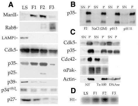
Fig. 1. (A) Western blots showing the distribution of several proteins in the low speed supernatant (LS) and Golgi fractions. Note the presence of Cdk5 and p35 in the F1 and F2 fractions. (B) Western blot showing that p35 remains associated with the F2 fraction after treatment with 2 M NaCl or 0.5 M Na2CO3 (pH 5 or 11). (C) Western blots showing that Cdk5 and p35 remain associated with the F2 fraction after treatment with 2% Triton X-100; in contrast, a DNase treatment releases Cdk5-p35 from the pellet. The Cdk5-p35 kinase present in the F1 fraction behaves similarly (not shown). Abbreviations: NT (non-treated), SN (supernatant), P (pellet). (D) Cdk5 histone H1 kinase activity in p35 IPs obtained from the low speed supernatant (LS), and Golgi fractions. For all experiments 30 µg of total protein were loaded in each lane.

An official website of the United States government
Here's how you know
Official websites use .gov
A
.gov website belongs to an official
government organization in the United States.
Secure .gov websites use HTTPS
A lock (
) or https:// means you've safely
connected to the .gov website. Share sensitive
information only on official, secure websites.
