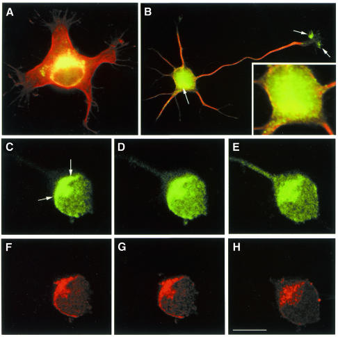
Fig. 2. p35 localizes to the Golgi apparatus in developing neurons. (A and B) Red-green overlays of digitized images showing the distribution of tubulin (red) and p35 (green) in cultured hippocampal pyramidal neurons. The cells were fixed prior to detergent extraction with 4% paraformaldehyde–0.12 M sucrose 1 day after plating and processed for immunofluorescence with antibodies against tyrosinated a-tubulin and p35. Note that a high p35 fluorescence signal is detected in a region of the cell body located in close apposition to the nucleus (arrow) and in the axonal growth cone (arrows). The insert in (B) shows a high magnification view of the cell body area. (C–H) A series of confocal images showing the distribution of p35 (C–E) and mannosidase II (F–H) in the cell body of a cultured hippocampal pyramidal neuron. Note the colocalization of both proteins in the region of the Golgi apparatus (arrows). Calibration bar: 10 µm.

An official website of the United States government
Here's how you know
Official websites use .gov
A
.gov website belongs to an official
government organization in the United States.
Secure .gov websites use HTTPS
A lock (
) or https:// means you've safely
connected to the .gov website. Share sensitive
information only on official, secure websites.
