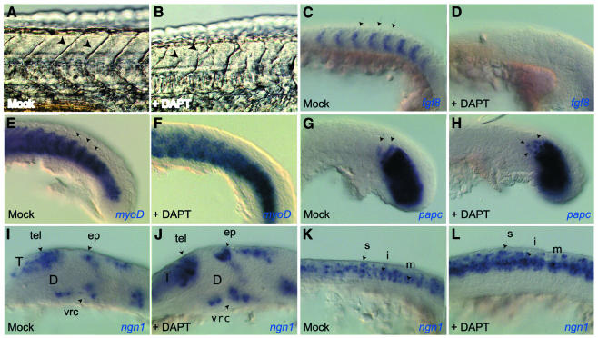
Fig. 2. DAPT affects somitogenesis (A–H) and neurogenesis (I–L) in vivo in a manner indistinguishable from Notch signaling deficiencies. Mock-treated (A, C, E, G, I, K) and DAPT-treated (B, D, F, H, J, L) embryos visualized at 24 h [(A) and (B), live views] or at 15 somites (C–L), flat mounts following whole-mount in situ hybridization with the probes indicated, anterior to the left and dorsal up except in (E) and (F) (dorsal views). (A–H), (K) and (L) are close-up views of the trunk and tail; (I) and (J) are close-up views of the brain. DAPT treatment alters somitic borders [arrows in (A) and (B)]. It affects AP polarization of the somitic mesoderm, normally revealed by the complementary expression of fgf8 and myoD [arrows in (C) and (E)], and randomizes expression of cycling-dependent genes such as papc in nascent somites [arrows in (G) and (H)]. In the embryonic nervous system, DAPT triggers a neurogenic phenotype, with an increased number of ngn1-positive neuroblasts in every proneural cluster [compare the clusters identified by arrows in (I) and (J), (K) and (L)]. D, presumptive diencephalon; ep, epiphyseal cluster; i, intermediate neurons; m, motoneurons; s, sensory neurons; T, presumptive telencephalon; tel, telencephalic cluster; vrc, ventro-rostral cluster.

An official website of the United States government
Here's how you know
Official websites use .gov
A
.gov website belongs to an official
government organization in the United States.
Secure .gov websites use HTTPS
A lock (
) or https:// means you've safely
connected to the .gov website. Share sensitive
information only on official, secure websites.
