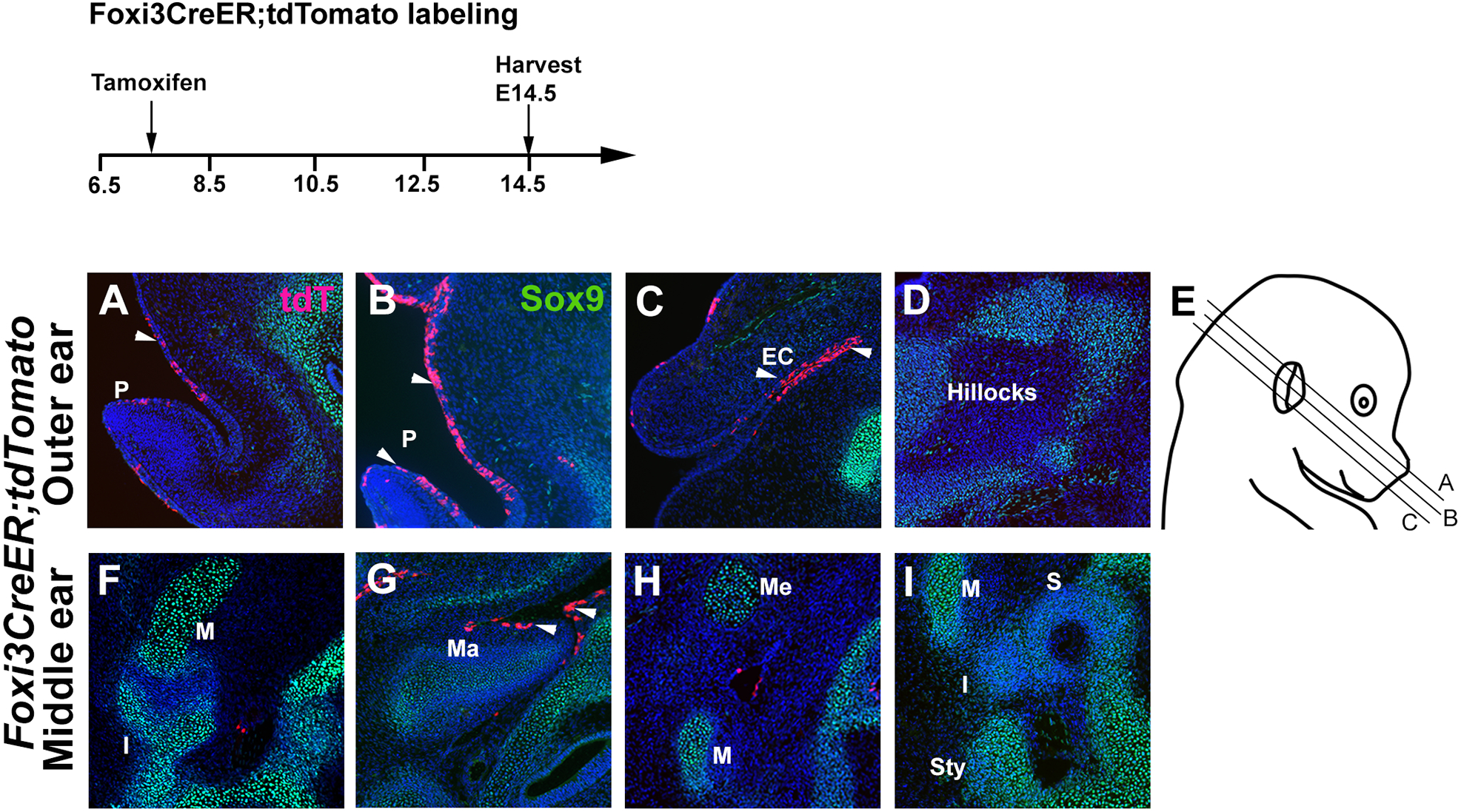
Figure 3: Foxi3-expressing cells contribute to the external and middle ear, but not the middle ear ossicles.

Foxi3CreER mice were mated with ROSA-Ai9 Cre reporter mice and the pregnant females received a single dose of tamoxifen at 7.5 dpc. Mice were analyzed for tdTomato expression at E14.5, together with Sox9 to show developing cartilage. (A-C) Foxi3 lineage-labeled derivatives can be seen in the ectoderm of the external ear pinna (P) and ear canal (EC), but not the mesenchymal auricular hillocks that label with Sox9 (D). (F-I) Although some cells can be observed in the endoderm of the Eustachian tube, the middle ear ossicles – the malleus (M), incus (I) and stapes (S) – are all unlabeled, as are Meckel’s cartilage (Me) and the styloid process (Sty)
