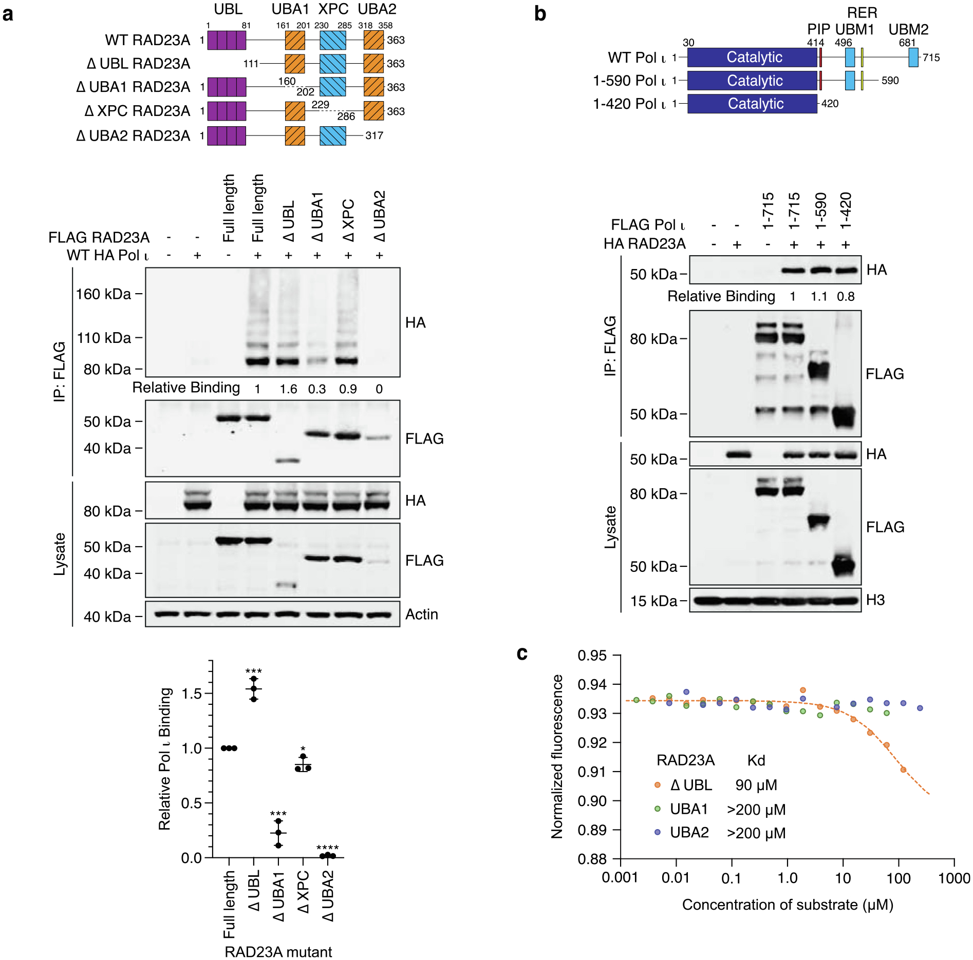
Figure 2.

The Pol ι catalytic domain binds UBA1 and UBA2 of RAD23A. (a) Immunoprecipitation of the RAD23A mutants illustrated in the schematic, from 293 T cells co-expressing WT HA Pol ι. Eluent and WCL (input) were immunoblotted as indicated. Relative binding was calculated based on the ratio of HA to FLAG proteins in the eluent. The bar graph represents quantification of relative binding from three repeats. Error bars represent standard deviation. (b) Immunoprecipitation of the indicated Pol ι mutants from HEK293T cells co-expressing WT HA RAD23A. Eluent and WCL (input) were immunoblotted as indicated. Relative binding was calculated based on the ratio of HA to FLAG proteins in the eluent. (c) Microscale thermophoresis analysis between the catalytic domain of Pol ι and the indicated fragments of RAD23A.
