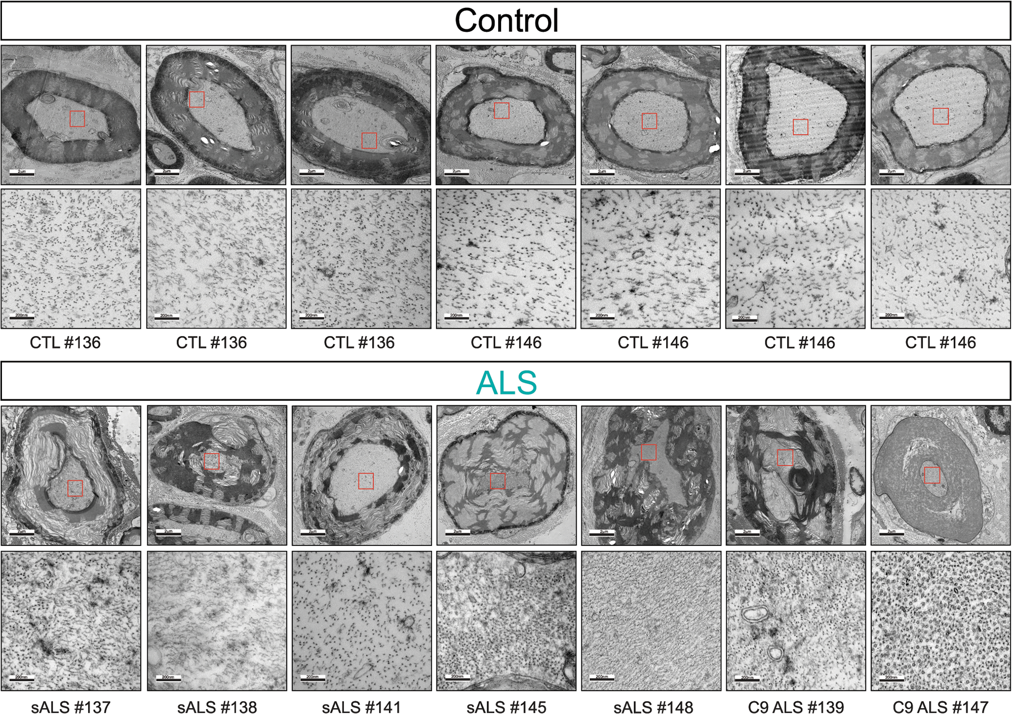
Extended Data Figure 4: Axonal shrinkage, collapse of neurofilament spacing, and tearing of myelin in sporadic ALS and C9-ALS.

Representative electron microscopy images of large caliber axon cross sections in the motor roots of postmortem human samples from healthy controls n = 2 (upper panel) and sporadic ALS (sALS, n=5) and C9orf72 ALS patients (C9 ALS, n=2) (lower panel). Increased magnification micrographs of the axoplasm showing altered spacing between neurofilament filaments is shown.
