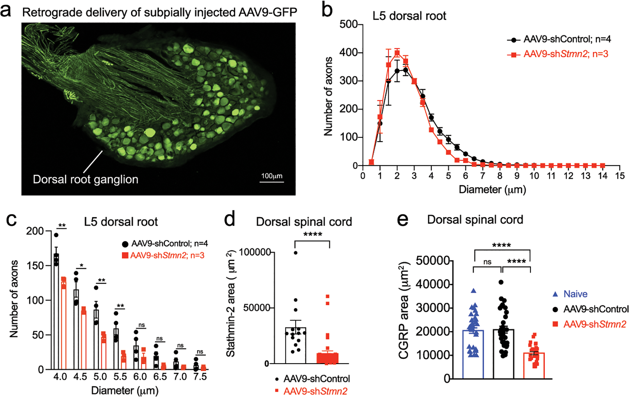
Extended Data Figure 5. Reduced stathmin-2 levels by subpial injection alters sensory marker in lumbar spinal cord:

(a) Representative image of lumbar dorsal root ganglion 2 months after subpial injection into lumbar spinal cord and subsequent retrograde delivery of AAV9 expressing green fluorescent protein (GFP). (b-c) Size distribution of axonal diameter of sensory axons innervating the dorsal spinal cord (b), and axon numbers in the 4 μm to 8 μm diameter range in the L5 dorsal root (c). Statistical analysis by two-sided, two-way ANOVA and Sidak’s multiple comparison test. P values range from P = 0.0189 to P = 0.0028. N=4 animals with AAV9-shControl and n=3 animals with AAV9-shStmn2. (d-e) Quantification related to Figure 4i,k of positive area for stathmin-2 (n=2 animals injected with AAV9-shControl and n=4 animals injected with AAV9-shStmn2, (P <0.0001) (d), and CGRP (e) in the dorsal spinal cord of age-matched non-injected naïve animals (n=5) or 8 months after subpial injection of AAV9 encoding either non-targeting sequence (n=4) or Stmn2 shRNA (n=4). P <0.0001. Statistical analysis by two-sided, Mann Whitney test (d) and Kruskal-Wallis nonparametric tests (e). All panels: Each data point represents an individual mouse. Error bars plotted as SEM. ****, P <0.0001; ***, P < 0.001; **, P < 0.01; *, P <0.05; ns, P >0.05.
