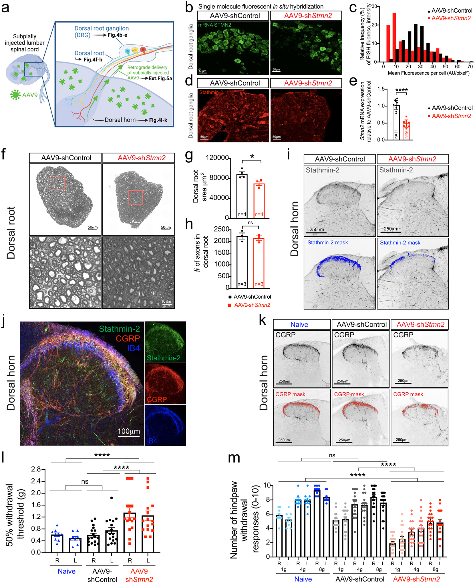
Figure 4: Reduced stathmin-2 levels in lumbar dorsal root ganglia impair hindlimb sensory system.

(a) Schematic of strategy to determine the impact of subpially injected AAV9-encoded shRNAs on the neurons in the dorsal root ganglion (DRG) innervating the dorsal spinal cord. Figure created using Biorender. (b-d) Representative confocal images from at least 3 animals per condition showing Stmn2 mRNA levels by single-molecule FISH (green, b) and its fluorescence distribution (c), and stathmin-2 protein (red, d) in the lumbar DRGs 8-months post-administration of AAV9-shRNAs. (e) Quantification of Stmn2 mRNA levels in DRG at 8-months post-injection, normalized to Gapdh. Statistics by two-sided, unpaired t-tests (P < 0.0001). (f-h) Representative images of cross-sectioned dorsal roots and higher magnification images showing axonal morphology and diameter size (f), quantification of area (g), and total number of sensory axons (h), of WT mice 8 months post-injection. Statistics by two-sided, unpaired t-tests (P = 0.0142). (i) Representative dorsal horns of lumbar spinal cord sections immunolabeled with stathmin-2, highlighted in blue, 8 months post-injection. N=2 animals on AAV9-shControl and n=4 animals with AAV9-shStmn2 were imaged with similar results. (j) Representative confocal micrograph of WT dorsal horn immunostained with stathmin-2 (green), CGRP (red), and IB4 (blue). N=3 wildtype animals were immunostained with similar results. (k) Representative lumbar spinal cord dorsal horn areas immunolabeled with CGRP, highlighted in red. N=5 non-injected (naïve) 20-month-old mice, or n=4 mice 8-months after subpial administration of AAV9-shRNAs were imaged with similar results. (l-m) Quantification of the 50% withdrawal threshold upon von Frey filament-based mechanical stimuli on mice hindlimbs (m) Quantification of hind paw response to increasing von Frey filament force stimuli on mice hindlimbs (l-m) Assays performed at 20 months-of-age when non-injected (naïve; n=9), or 8-months post-administration of AAV9-shControl (n=17) and AAV9-shStmn2 (n=16). Statistics by two-sided, one-way ANOVA Kruskal-Wallis with Dunn’s multiple comparisons test. P < 0.0001. For p values between specific conditions please see Source Data for Figure 4. All panels: Each data point represents an individual mouse. Error bars plotted as SEM. ****, P <0.0001; ***, P < 0.001; **, P < 0.01; *, P <0.05; ns, P >0.05.
