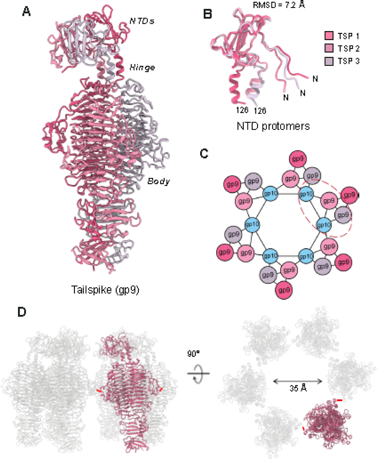
Figure 4. The asymmetric structure of the P22 tailspike.

(A) The structure of the full-length P22 tailspike reveals an asymmetric NTD leaning to the left, connected to a triple β-helix body. (B) Superimposition of the tailspike N-termini (res 1–126). (C) Schematic diagram of the symmetry mismatch between the tail hub and trimer tailspikes. The red dashed line circles the pseudo-2:1 binding interface. (D) Quaternary structure of P22 tailspikes observed in the MV. Six tailspike (one subunit in red and five in light gray) are shown in a side view (left panel) and rotated along the y-axis by 90 degrees (right panel). The red dashes denote the smallest distance between adjacent tailspike subunits.
