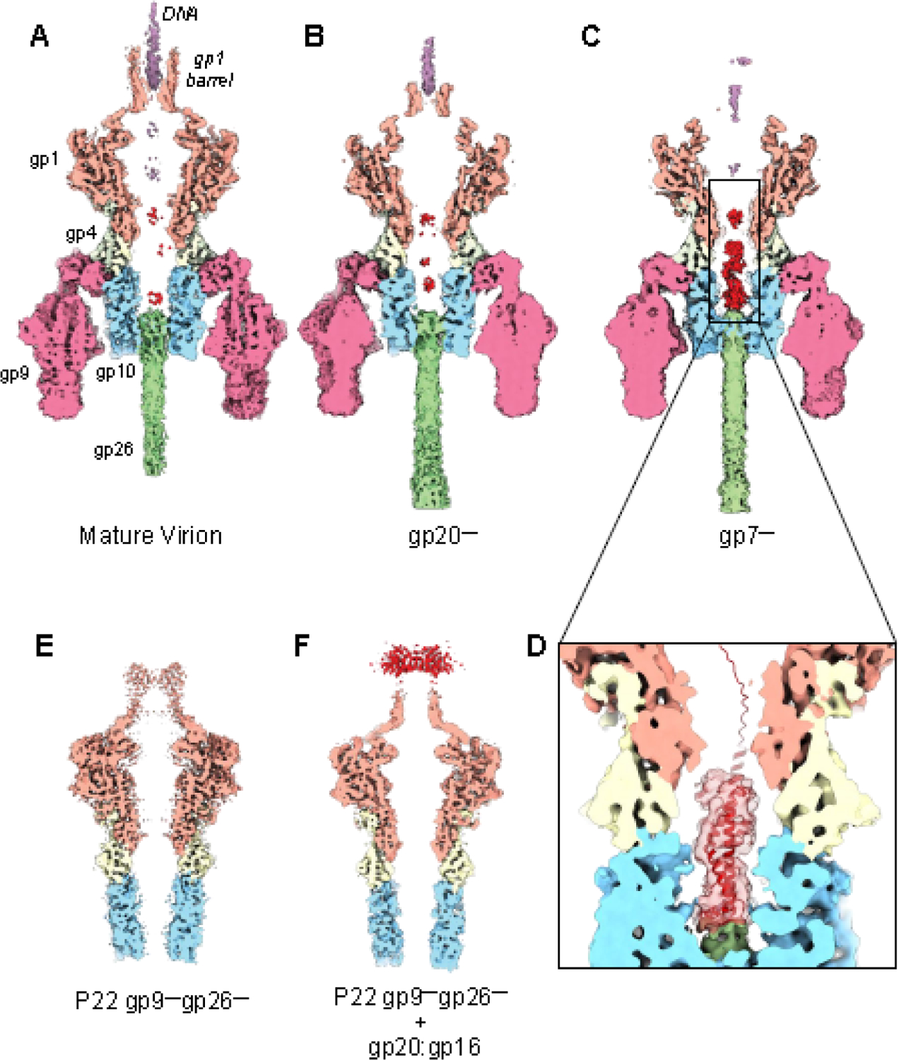
Figure 5. Cryo-EM reconstructions of P22 virion mutants.

Cross-sectional views of P22 tail asymmetric reconstructions of the (A) P22 MV and mutants lacking one ejection protein, (B) P22 gp20— and (C) P22 gp7—. (D) A magnified view of the density in the channel (in light red) overlaid to an AlphaFold model of gp20. (E) Cross-sectional view of a P22 mutant lacking the tailspike and tail needle (P22 gp9—/gp26—). (F) Cross-sectional view of P22 gp9—/gp26— incubated with an excess of recombinant gp20:gp16 complex purified from a soluble bacterial lysate. All the maps shown in this figure were sharpened, low pass filtered to 5.2 Å, and are displayed at 2.0 σ.
