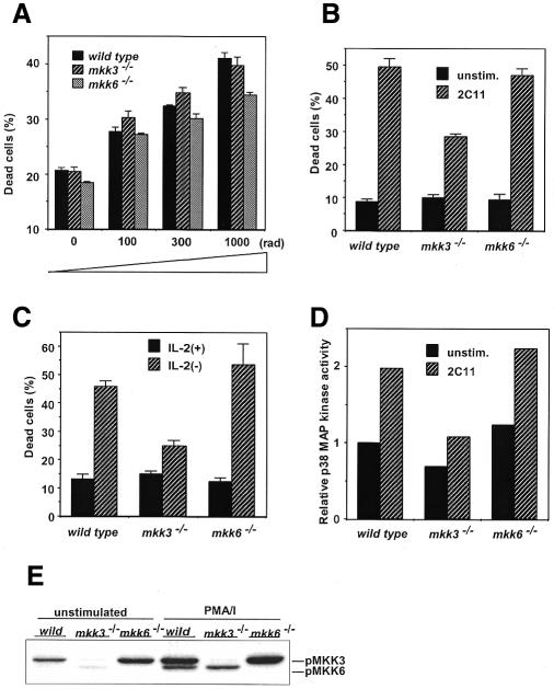
Fig. 4. Differential susceptibility to apoptosis between CD4+T cells from Mkk6–/– and Mkk3–/– mice. (A) Freshly prepared CD4+T cells are γ-irradiated. (B and C) CD4+T cells were stimulated with anti-CD3/CD28 mAbs for 3 days. Cells are restimulated by anti-CD3 mAb (B) or cultured in the absence of IL-2 (C) for 20 h and analyzed for viability. (D) Impaired p38MAPK activity in CD4+T cells from Mkk3–/– mouse. Cells were stimulated by anti-CD3 mAb for 90 min and determined for p38MAPK activity. (E) MKK3 and MKK6 activation by PMA/ionomycin (PMA/I) stimulation. Cells were stimulated in vitro for 15 min and examined for activated MKK3/6.

An official website of the United States government
Here's how you know
Official websites use .gov
A
.gov website belongs to an official
government organization in the United States.
Secure .gov websites use HTTPS
A lock (
) or https:// means you've safely
connected to the .gov website. Share sensitive
information only on official, secure websites.
