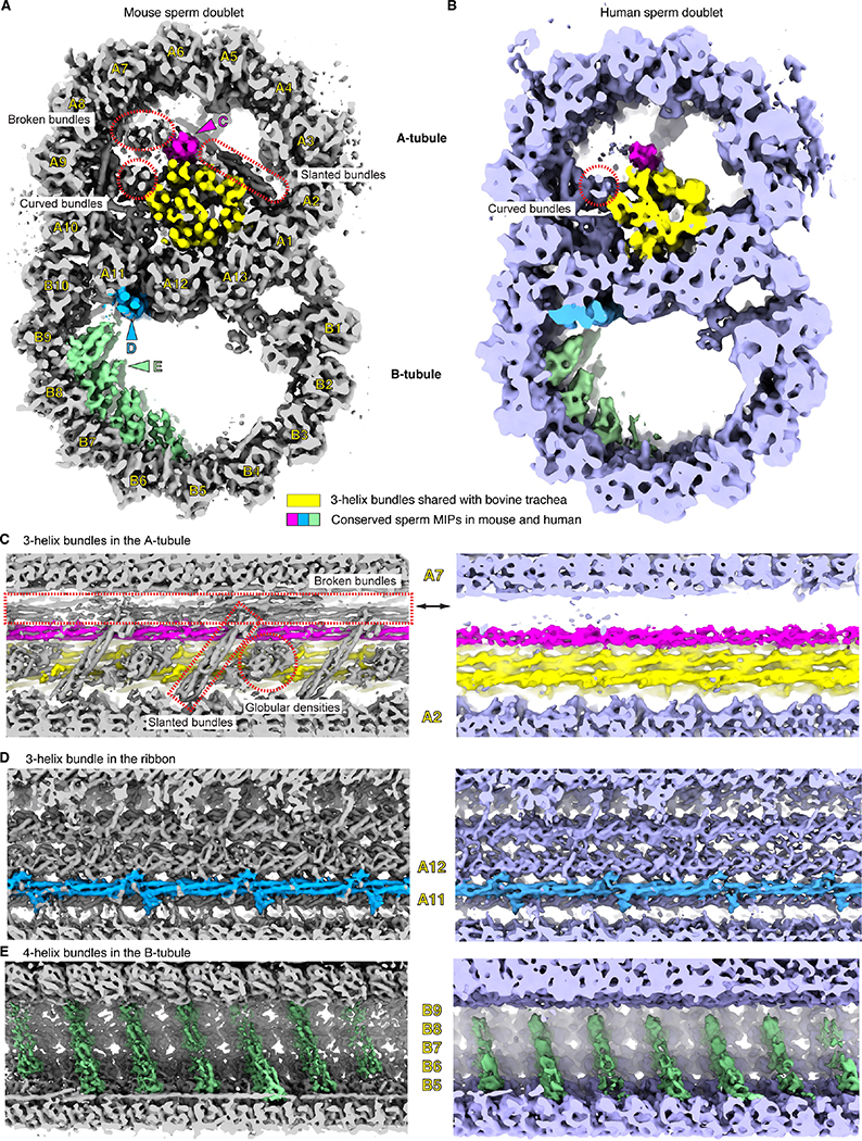
Figure 1. The 3D reconstructions of mouse and human sperm doublets revealed novel MIPs.
(A), (B) Transverse cross-section views of the doublets of mouse (A) and human (B) sperm. Conserved sperm MIP densities are highlighted (pink, blue and green) and the corresponding viewing angles of (C)-(E) are indicated (colored arrowheads). The 3-helix densities in A-tubule shared with Bovine trachea doublets (EMD-24664) are colored (yellow) 13. Divergent sperm densities are also indicated (red dashed shapes). Individual protofilaments of the doublets are labeled as A1–13 and B1–10. (C)-(E). Zoom-in views of the conserved sperm MIP densities along the longitudinal axis. In (C), mouse sperm-specific densities are indicated and labeled (red dashed shapes, see more in Figures S2 and S3). In (E), although the striations are 8 nm apart from one another, the overall periodicity is 48 nm.

