
Figure 5. PARKIN is dispensable for MLRO formation in prolonged cultured hepatocytes.
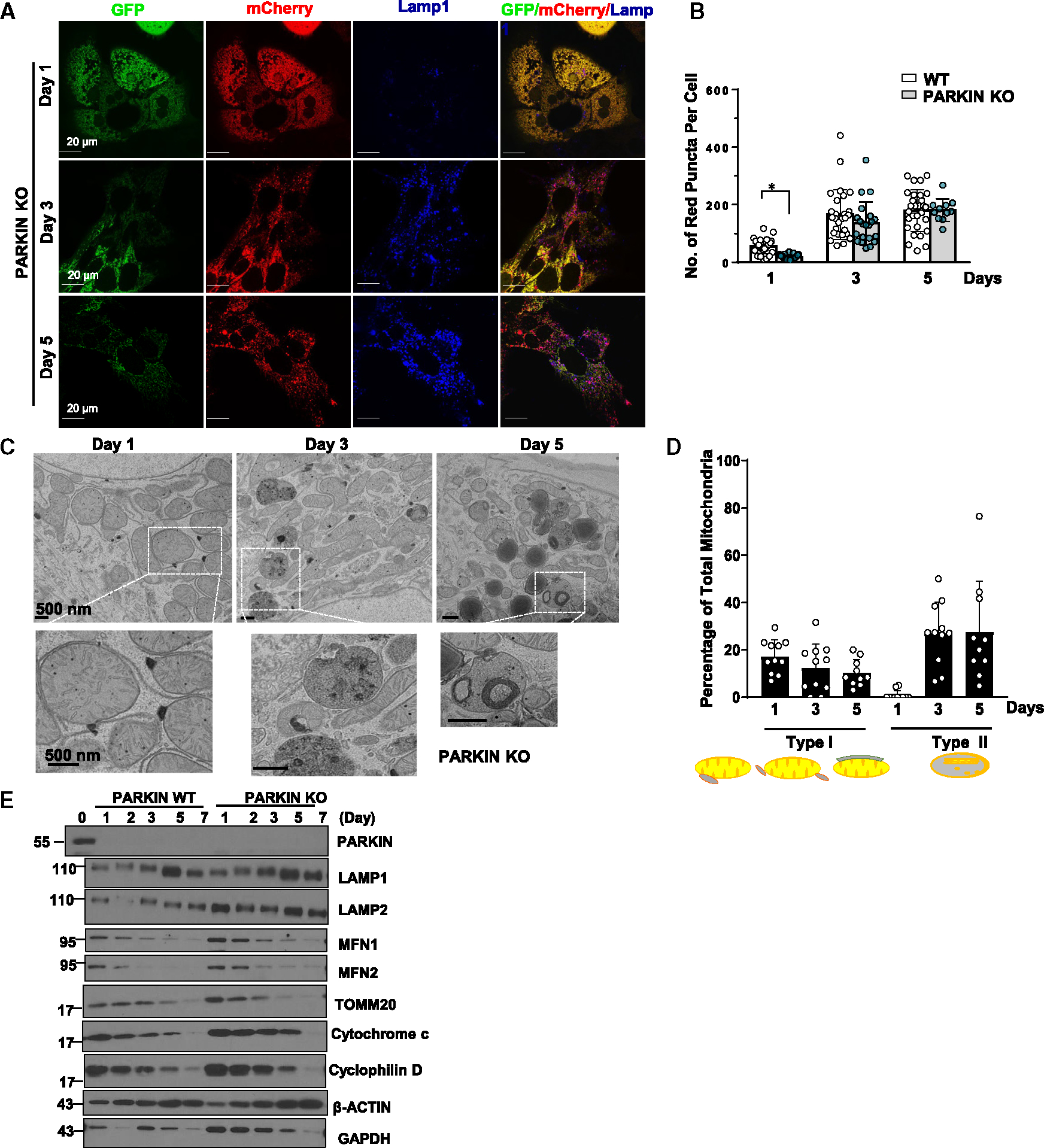
(A) PARKIN KO primary mouse hepatocytes were infected with adenovirus-Cox8-GFP-mCherry (MOI = 10) from seeding for overnight. Cells grown on coverslips were fixed in 4% PFA at indicated time points followed by LAMP1 immunofluorescence staining. Images were captured using confocal microscopy.
(B) Quantification data for the number of red-only mitochondria in each cell from Cox8-GFP-mCherry assay. Data are presented as mean ± SD. At least 15 fields from two independent mouse hepatocytes perfusion were quantified.
(C) Day 1 to day 5 cultured PARKIN KO mouse hepatocytes were examined using transmission electron microscopy (TEM).
(D) Type I and type II MLRO structure were quantified from (C) and normalized by the total mitochondria number in each field.
(E) WT and PARKIN KO primary mouse hepatocytes were cultured for indicated times. Total cell lysates were subjected to western blot analysis. *p < 0.05, two-tailed Student’s t test.