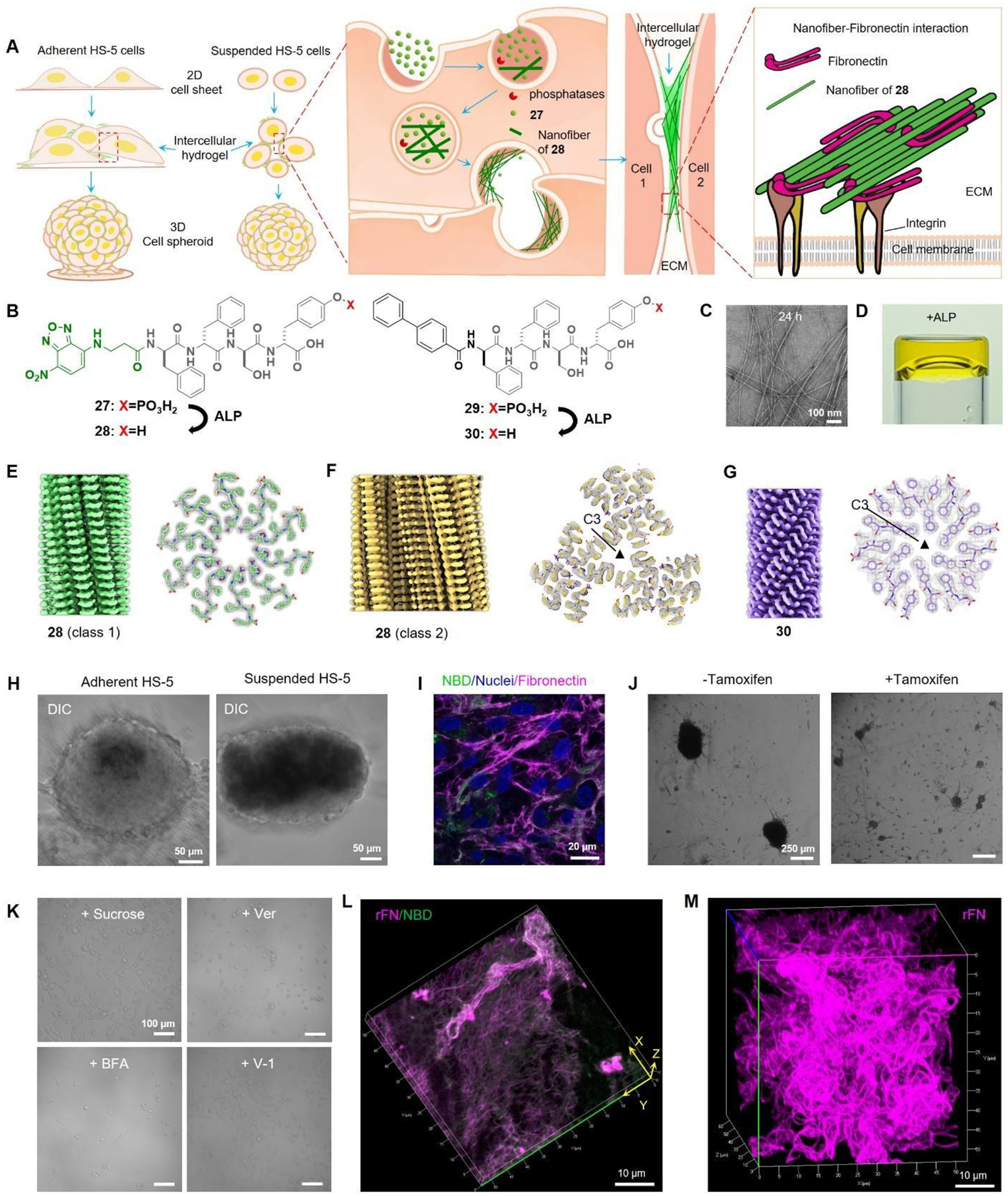
Figure 11.

(A) Illustration of the transcytotic dephosphorylation of 27 forming intercellular hydrogels that colocalize with fibronectin to enable spheroids. (B) Structures of 27, 28 and 29. (C) TEM images of 27. (D) ALP converts 27 to gel. 3D reconstruction and cross section of (E) 28 (class 1), (F) 28 (class 2), and (G) 29 filaments. (H) Adherent or suspended HS-5 cells treated with 27 to form spheroids. (I) Immunofluorescence staining of fibronectin in HS-5 cells treated with 27. (J) Suspended TKO cells with or without tamoxifen knockout incubated with 27. (K) Suspended HS-5 cells under different conditions (27 at 200 μM): + Sucrose (sucrose pre-treated cells + 27); + Ver (27 + 50 μM Verapamil); + BFA (27 + 300 nM Brefeldin A); + V-1 (27 + 10 μM Vacuolin-1). (L) 3D rendering of 27 treated with ALP and then incubated with rhodamine-fibronectin (rFN). (M) 3D rendering of 29 treated with ALP and then incubated with rFN. Adapted from Ref.67 Copyright Springer 2023.
