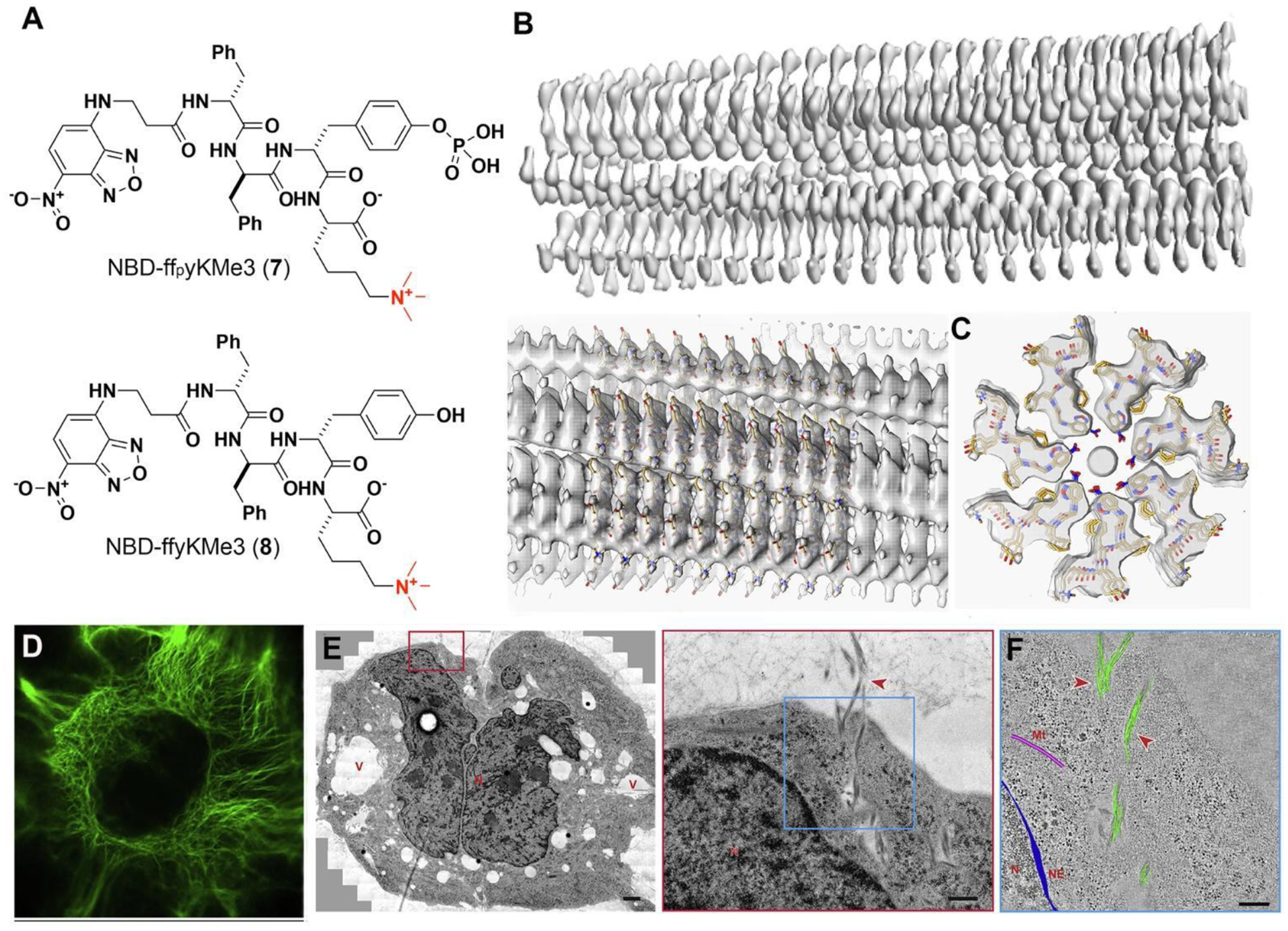
Figure 5.

(A) Structures of 7 and 8. (B) 3D cryo-EM reconstruction of the filaments of 8. (C) Top views of the cross-section of the EM density of the filament and the stick representation of the peptides. (D) CLSM images of Saos-2 cells treated with 7. Scale bars, 10 mm. (E) TEM image treated Saos-2 cell (7, 200 μM, 24 h) and higher-magnification electron micrograph of the red boxed area. (F) 3D reconstruction models of the filament bundles (green), microtubules (pink), and nuclear envelope (blue) on an electron tomographic image of the blue boxed area in (E). Adapted from Ref.44. Copyright of the authors 2020 CC BY-NC-ND.
