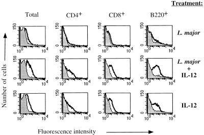
FIG. 3.
IL-12 binding to LN cells from BALB/c mice given exogenous IL-12. Shown is a two-color flow-cytometric analysis of IL-12 binding to lymphocyte subsets using LN cells from BALB/c mice infected 2 days previously, BALB/c mice infected 2 days previously and treated in vivo with 0.2 μg of IL-12 at the time of infection, and BALB/c mice treated with 0.2 μg of IL-12 alone 2 days prior to analysis. The peaks outlined by the dark solid line and the shaded peaks are as defined in the legend for Fig. 1. These results are representative of three experiments.

