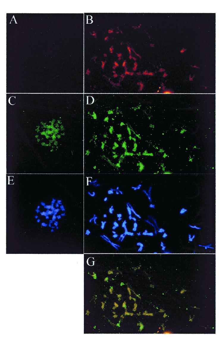
Fig. 1. Localization of EBP2 and EBNA1 on mitotic chromosomes. Mitotic chromosome spreads are shown from Raji (right panels) and BL41 (left panels) Burkitt’s lymphoma cells that do and do not express EBNA1, respectively. (A) and (B) EBNA1 staining with monoclonal antibody. (C) and (D) EBP2 staining with rabbit polyclonal antibody. (E) and (F) DAPI staining. (G) Overlay of images (B) and (D) showing co-localization (yellow). Images were captured at 630-fold magnification.
