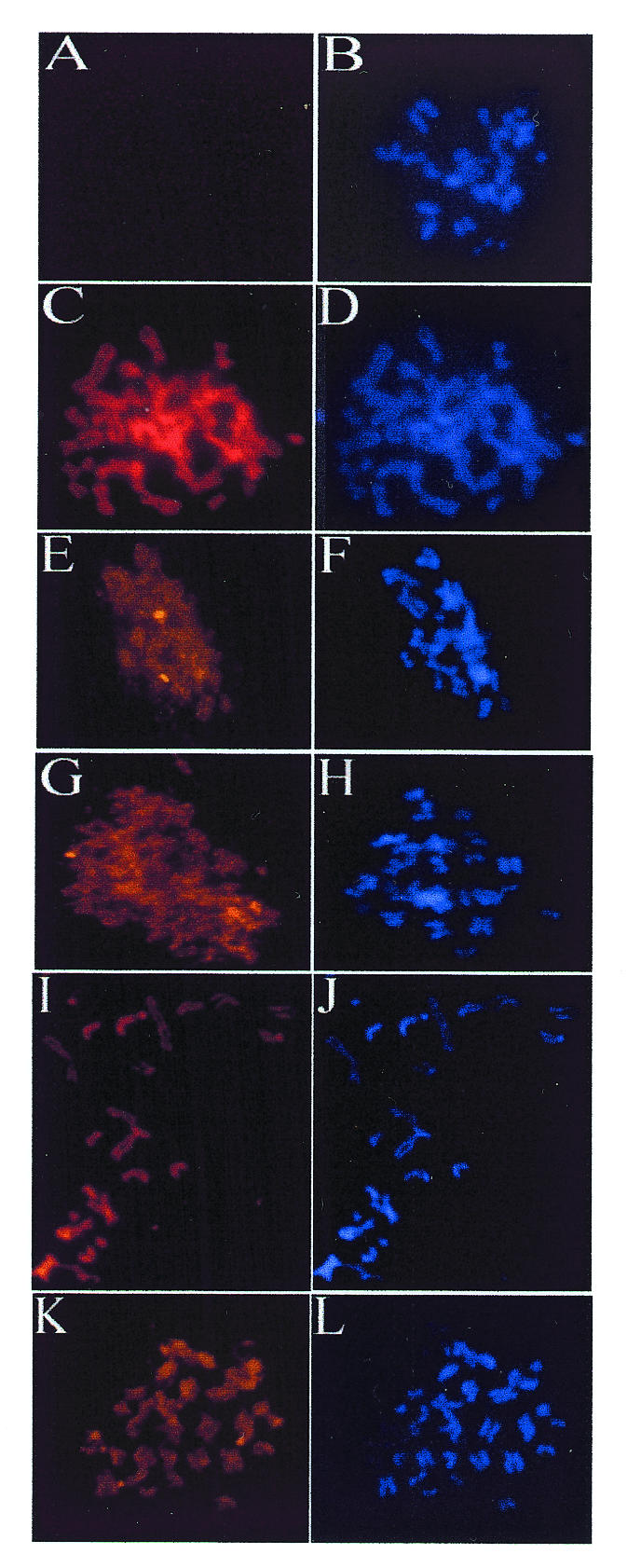
Fig. 4. Mitotic localization of EBNA1 mutants. C33A cells that are EBNA1-negative (A and B) or that express EBNA1 (C and D), Δ325–376 (E–H), Δ356–362 (I and J) or Δ367–376 (K and L) were blocked in mitosis by colcemid treatment, and chromosomes were spread for microscopy. EBNA1 proteins were detected with monoclonal antibody (left panels), and DNA was detected with DAPI (right panels). Images in left panels were captured using similar exposure times and 630-fold magnification.
