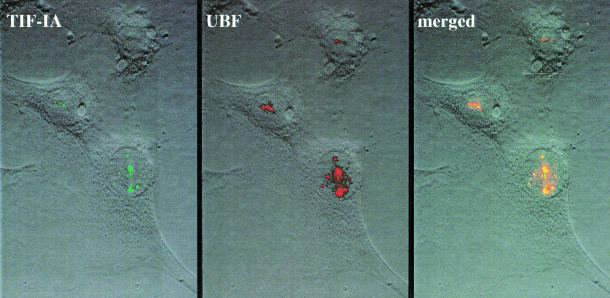
Fig. 2. Nucleolar localization of TIF-IA. NIH 3T3 cells were transfected with CMV-FLAG-hTIF-IA, fixed in methanol for 1 min at –20°C, washed once with –20°C acetone and several times with phosphate-buffered saline. TIF-IA was visualized by indirect immunofluorescence using M2 antibodies (Sigma, 1:200) and fluorescein isothiocyanate-conjugated goat anti-mouse IgGs (Dianova, 1:300). UBF was stained with anti-UBF serum (1:500) and Texas red-conjugated goat anti-human IgGs (Dianova, 1:200).

An official website of the United States government
Here's how you know
Official websites use .gov
A
.gov website belongs to an official
government organization in the United States.
Secure .gov websites use HTTPS
A lock (
) or https:// means you've safely
connected to the .gov website. Share sensitive
information only on official, secure websites.
