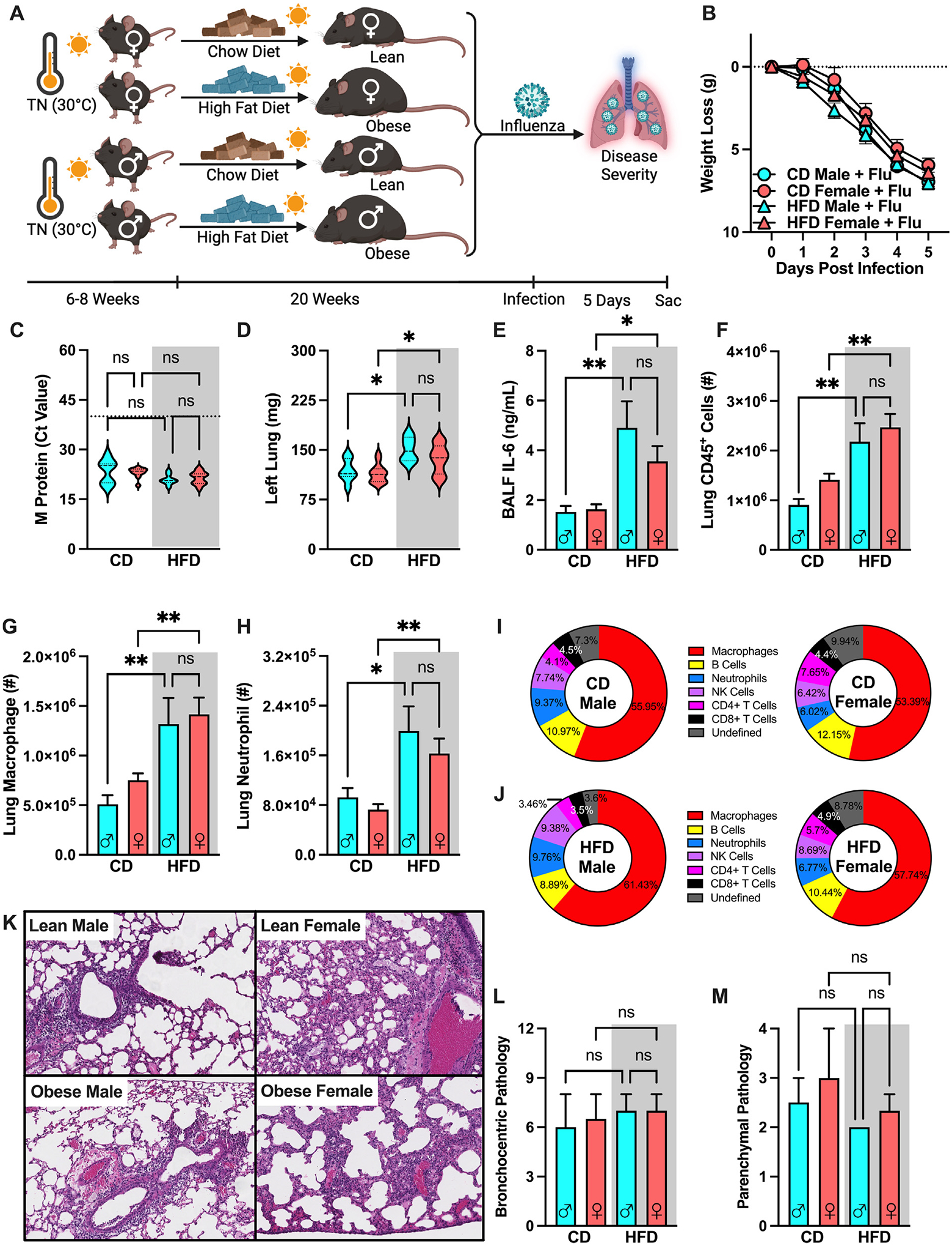
Fig. 6.

Obese male and female mice have comparably exacerbated obesity-associated influenza disease severity. (A) C57BL/6 male female mice (n = 6–15/group; combined results of 4 independent experiments) were placed in TN housing and fed CD or HFD for 20 weeks before intranasal challenge with influenza virus (30HA Units). (B) Weight loss post-infection. (C) Lung M protein expression quantified via qPCR. (D) Left lung weight 5 days post-infection (E) BALF IL-6 levels quantified via cytokine ELISA. (F) Total lung CD45+ immune cell infiltration, measured by flow cytometry. (G) Lung macrophage absolute numbers (CD11bhiF4/80hi). (H) Lung Neutrophil absolute numbers (CD11bhiGR1hi). (I) Infected immune cell population percentage of total lung CD45+ cells in TN-housed CD-fed males and females. (J) Infected immune cell population percentage of total lung CD45+ cells in TN-housed HFD-fed males and females. (K) Histopathological score of bronchocentric pneumonia (L) Histopathological score of inflammatory extension to parenchyma. (M) Representative H&E staining of unflushed left lungs (light microscope, 10X). Means ± SEM. Student’s t test or one-way analysis of variance; **p < 0.01, ***p < 0.001, ****p < 0.0001. BALF = bronchoalveolar lavage fluid; CD = chow diet; ELISA = enzyme-linked immunosorbent assay; H&E = hematoxylin & eosin; HFD = high-fat diet; IL-6 = interleukin-6; SEM = standard error of the mean; TN = thermoneutral.
