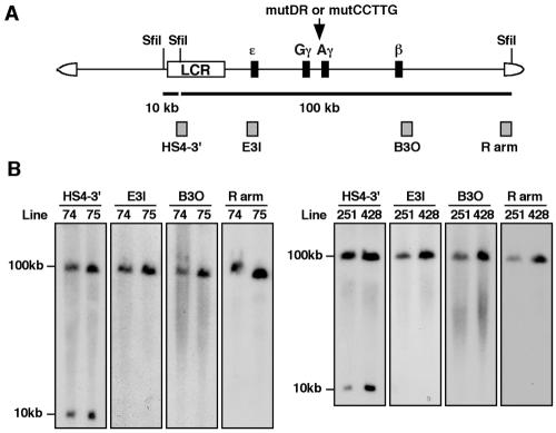
FIG. 2.
Structural analysis of human β-globin YAC TgM. (A) Schematic representation of the human β-globin YAC. The positions of the β-like globin genes are shown relative to the LCR. SfiI restriction enzyme sites are indicated as vertical lines. Probes (hatched boxes) used for long-range structural analysis and anticipated restriction enzyme fragments after SfiI digestion are shown with their sizes (solid thick lines). (B) Long-range transgene analysis of mutDR (left panel) and mutCCTTG (right panel) YAC transgenic mice. The whole β-globin locus is contained within two SfiI fragments (10 and 100 kb, as in panel A). DNA from thymus cells of transgenic mice was digested with SfiI in agarose plugs, separated by pulsed-field gel electrophoresis, and hybridized separately to probes (indicated on the top of each panel) from the β-globin locus or the right YAC vector arm. The sizes of the expected bands are shown on the left.

