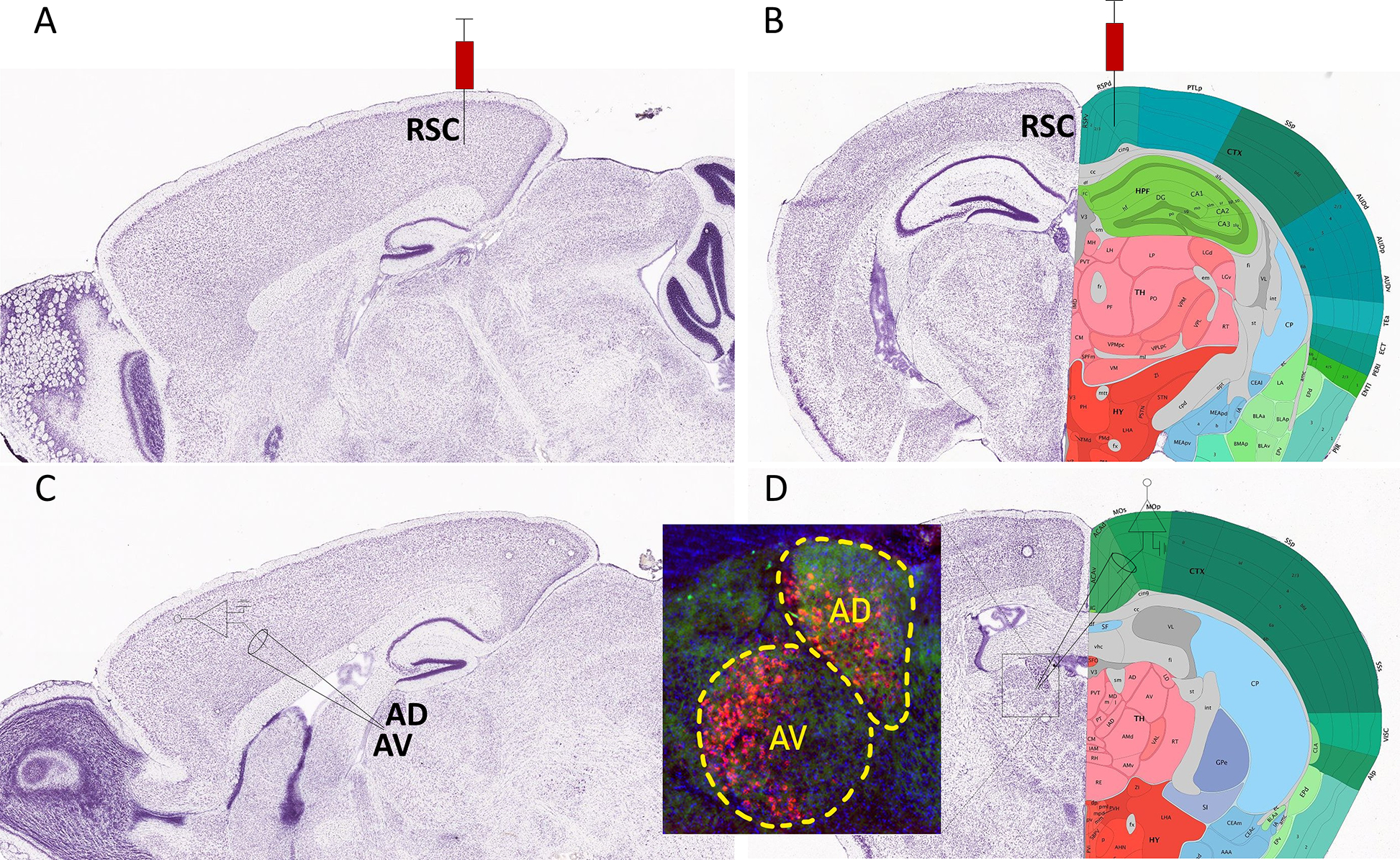
Figure 3. Schematic representation of retrobead injection into the retrosplenial cortex to label neurons in the anterodorsal (AD) and anteroventral (AV) thalamic nuclei.

A) Parasagittal image of a Nissl stained section (image #18) from the Allen Reference Atlas – Mouse Brain (postnatal day 56) illustrating the site of red retrobead injection in the retrosplenial cortex (RSC). B) Nissl stained image with anatomical annotations of a coronal section (image #75) from the same atlas also showing the site of injection in the RSC. C) Parasagittal image of a Nissl stained section (image #16) from the same atlas illustrating the site of electrophysiological recordings in the AD and AV thalamic nuclei. D) Nissl-stained image with anatomical annotations of a coronal section (image #62) from the same atlas also showing the site of electrophysiological recordings in the AD and AV thalamic nuclei. The inset shows an image of red retrobeads in the AD and AV nuclei from a mouse that received an injection in the RSC. Allen Mouse Brain Atlas, mouse.brain-map.org.
