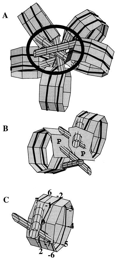
Figure 9.
(A) Graphic presentation of a pentanucleosome according to one geometrical solution for the 30 nm fibre (26). The black circle depicts the area of the fibre where DNase I cannot digest the nucleosomal and linker DNA because of the steric hindrances imposed. (B) Two adjacent nucleosomes from (A) shown in a different projection. Polygons P depict the corresponding protected areas. (C) A nucleosome, showing the numbering of the digestion sites.

