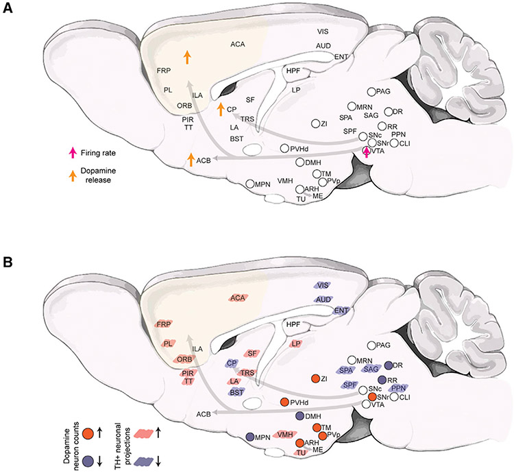
Figure 1. Brain-wide impact of ketamine exposure on the dopaminergic modulatory system.
A) Schematic summary of currently known alterations in dopaminergic neuronal activity and dopamine release after ketamine exposure.16,17
B) Schematic summary (this study) of the brain-wide alterations in the DA system after 10 days of repeated ketamine (30 and 100 mg/kg) exposure. Up and down arrows indicate increase and decrease, respectively. Note that, for the 30 mg/kg ketamine treatment, statistically significant decreases in TH+ neuron counts were only observed in DR, DMH, and MPN and increase in ZI and SNr. The TH+ neuronal projections changes are only shown for the 100 mg/kg (10 days) treatment. Abbreviations used are standard Allen Brain Atlas terms, as follows: ACA, anterior cingulate area; ACB, nucleus accumbens; ARH, arcuate hypothalamic nucleus; AUD, auditory areas; BST, bed nuclei of the stria terminalis; CLI, central linear nucleus raphe; CP, caudoputamen; DMH, dorsomedial nucleus of the hypothalamus; DR, dorsal nucleus raphe; ENT, entorhinal area; FRP, frontal pole, cerebral cortex; HPF, hippocampal formation; IF, interfascicular nucleus raphe; ILA, infralimbic area; LA, lateral amygdalar nucleus; LP, lateral posterior nucleus of the thalamus; MPN, medial preoptic nucleus; MRN, midbrain reticular nucleus; ORB, orbital area; PAG, periaqueductal gray; PIR, piriform area; PL, prelimbic area; PPN, pedunculopontine nucleus; PVH, paraventricular hypothalamic nucleus; PVp, paraventricular hypothalamic nucleus, posterior part; RR, midbrain reticular nucleus, retrorubral area; SAG, nucleus sagulum; SF, septofimbrial nucleus; SNc, substantia nigra, compact part; SPA, subparafascicular area; SPF, subparafascicular nucleus; TM, tuberomammillary nucleus; TRS, triangular nucleus of septum; TT, taenia tecta; TU, tuberal nucleus; VIS, visual areas; VMH, ventromedial hypothalamic nucleus; VTA, ventral tegmental area; ZI, zona incerta.

