
FIGURE 1.
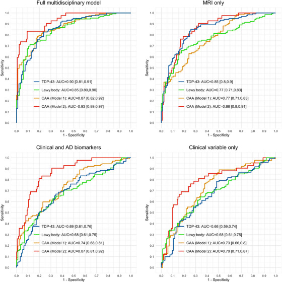
ROC analysis of comorbid non‐ADNC positivity prediction in autopsy‐confirmed main study cohort of CU elderly individuals, individuals with MCI, and individuals with dementia due to AD. The full multidisciplinary model used harmonized ICV‐adjusted regional volumetric estimates from automated MUSE parcellation, demographics, number of APOE alleles (ε2 and ε4 separately), MMSE, CDR, and end‐of‐life ADNC status (Aβ positivity and tau positivity separately) as primary predictor variables. Reference classifiers were constructed to assess the added value of ADNC biomarkers together with MRI volumetrics. TDP‐43 positivity: none versus inclusions in amygdala, hippocampus, entorhinal/inferior temporal cortex, or neocortex. LBD positivity: none versus amygdala predominant, limbic (transitional), or neocortical (diffuse) LBD pathology. CAA positivity: none or mild versus moderate or severe CAA in Model 1; none versus mild, moderate, or severe CAA in Model 2.