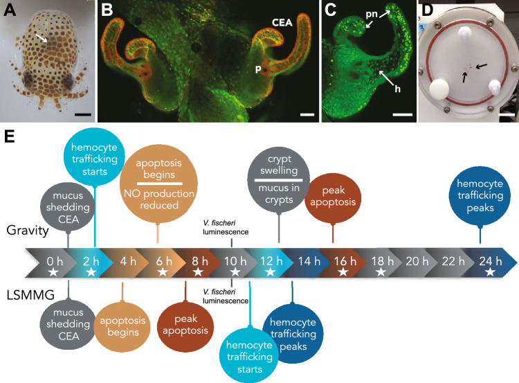
Figure 1.
Overview of the Euprymna scolopes and Vibrio fischeri symbiosis. (A) Hatchling E. scolopes with the location of the symbiotic light organ depicted by the white arrow. Bar, 500 µm. (B) Epifluorescent image of the nascent light organ stained with acridine orange visualizing the superficial ciliated epithelial appendages (CEA) that entrain bacteria into the vicinity of pores (p) on the surface of the light organ. Bar, 50 µm. (C) One-half of the light organ 16 h after inoculation with symbiosis-competent strains of V. fischeri. Acridine orange staining reveals the pattern of pycnotic nuclei (pn) typically associated with the bacteria-induced apoptotic cell death event. The symbiotic V. fischeri also triggers the migration of macrophage-like hemocytes (h) into the blood sinus underlying the CEA. Bar, 50 µm. (D) Example of a high aspect-ratio vessel (HARV) used to simulate low-shear modeled microgravity (LSMMG). Squid, indicated by the black arrows, are in “free fall” suspended in the middle of the HARV. Bar, 3 cm. (E) Timeline of bacteria-induced developmental events in the host under normal gravity conditions compared to low shear modeled microgravity conditions (LSMMG). Stars indicate time points targeted for NanoString gene expression assay.

