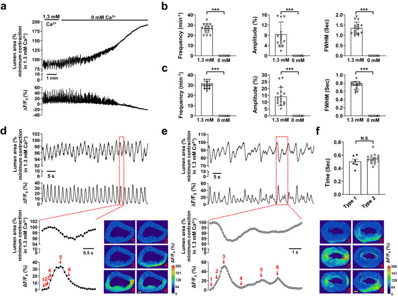
Fig. 1. Intercellular Ca2+ waves induce rhythmic contractions, leading to basal tone, in IAS slices.
a Ca2+ signals and associated contractions in the presence and absence of extracellular Ca2+. The summed area values were normalized to the maximal relaxation or minimal contraction during rhythmic contractions in the presence of 1.3 mM extracellular Ca2+ in this panel and throughout the other figures. b Summary of the effects of extracellular Ca2+ removal on contractions. c Summary of the effects of extracellular Ca2+ removal on Ca2+ signals. FWHM: Full Width at Half Maximum. Data from the last 30 s in the 0 mM Ca2+ medium were used to calculate the zero-calcium treatment. n = 16. d Type 1 intercellular calcium waves with regular amplitude and duration (lower trace) were temporally associated with rhythmic contractions (upper trace) in a one-to-one manner. Inset: a single event, indicated by the red dotted line box, is shown on an expanded scale. Images show changes in lumen area and Ca2+ fluorescence (ΔF/F0(%)); numbers in the images correspond to those marked near the ΔF/F0(%) trace, scale bar = 200 μm. e Type 2 intercellular calcium waves (lower trace) with irregular amplitude and duration were associated with rhythmic contractions (upper trace). Inset: a single event, indicated by the red dotted line box, is shown on an expanded scale. The images display temporal changes in the lumen area and Ca2+ fluorescence (ΔF/F0(%)); numbers in the images correspond to those marked near the ΔF/F0 trace, scale bar = 200 μm. f Delay of contraction onset relative to Ca2+ wave onset. n = 6 for type 1, n = 14 for type 2; N.S. no significant difference, ***p < 0.001, as determined by paired two-tailed (b, c) or unpaired two-tailed (f) Student’s t test.

