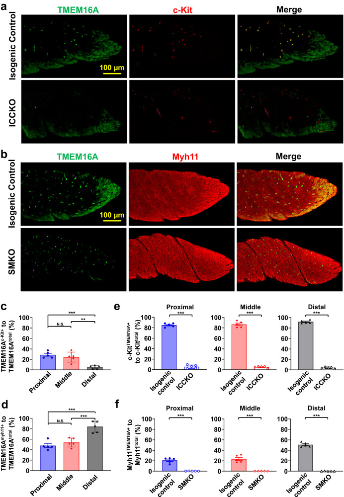
Fig. 5. Spatial pattern of TMEM16A expression in the IAS.
a Upper row: Immunostaining of TMEM16A (left) and c-kit protein (middle), and their colocalization (right) in a control IAS. Lower row: The same immunostaining in a TMEM16AICCKO IAS. Maximum intensity projections are shown from a 20-plane 3D stacked image in both a and b below. Note that TMEM16A immunostaining signals are absent in c-kit-positive interstitial cells of Cajal (ICC) in the TMEM16AICCKO IAS. b Upper row: Immunostaining of TMEM16A (left) and myh11 protein (middle), and their colocalization (right) in a control IAS. Lower row: The same immunostaining in a TMEM16ASMKO IAS. Note that TMEM16A immunostaining signals are absent in Myh11-expressing smooth muscle cells (SMCs) in the TMEM16ASMKO IAS. c Zonal quantification of TMEM16A-containing pixels in ICC relative to total TMEM16A-containing pixels using isogenic controls for TMEM16AICCKO mice (n = 5). Zones are defined as one-third segments of the IAS; see Supplementary Fig. 3 for illustration. N.S. indicates no statistical significance, **p < 0.01, ***p < 0.001, as determined by one-way ANOVA analysis with Tukey’s multiple comparisons test. d Zonal quantification of TMEM16A-containing pixels in SMCs relative to total TMEM16A- containing pixels using isogenic controls for TMEM16ASMKO mice (n = 5). N.S. indicates no statistical significance, ***p < 0.001, as determined by one-way ANOVA analysis with Tukey’s multiple comparisons test. e Efficiency of TMEM16A deletion in ICCs in three different zones: proximal, middle, and distal regions of the IAS in TMEM16AICCKO mice (n = 5). ***p < 0.001, as determined by unpaired two-tailed Student’s t test. f Efficiency of TMEM16A deletion in SMCs in three different zones: proximal, middle, and distal regions of the IAS in TMEM16ASMKO mice (n = 5). ***p < 0.001, as determined by unpaired two-tailed Student’s t test.

