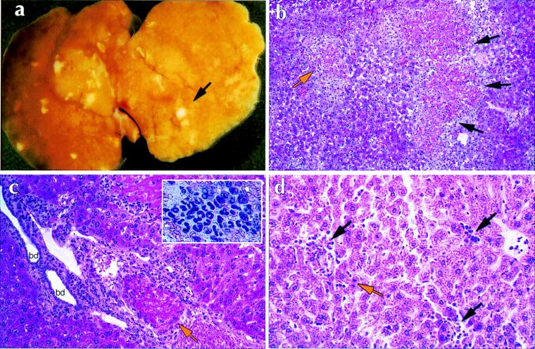
FIG. 5.
Histopathology of E. chaffeensis infection in C.B-17-scid/bg and C.B-17 mice. (a) Whole mount of a liver of a C.B-17-scid/bg mouse at 17 days postinfection. The arrow indicates an area of necrosis. (b to d) Hematoxylin-eosin staining of paraffin sections of infected livers. (b) C.B-17-scid/bg liver showing extensive areas of acidophilic coagulative necrosis of entire lobule (orange arrow) and confluent lobules (black arrows). Note the inflammatory infiltrate at the periphery of necrotic lobules. Magnification, ×100. (c) C.B-17-scid/bg liver showing portal tract with organizing thrombus in portal vein (orange arrow). There is a diffuse mononuclear inflammatory infiltrate in the portal tract penetrating the limiting plate and bordering hepatocytes. bd, bile ducts. Magnification, ×250. The inset shows a high-magnification view of a typical granulomatous infiltrate in a C.B-17-scid/bg liver. (d) Presence of lymphohistiocytic infiltrates in the liver of a C.B-17 mouse at 3 days postinfection (black arrows). Note apoptotic hepatocytes, characterized by condensed nuclei (orange arrow). Magnification, ×250.

