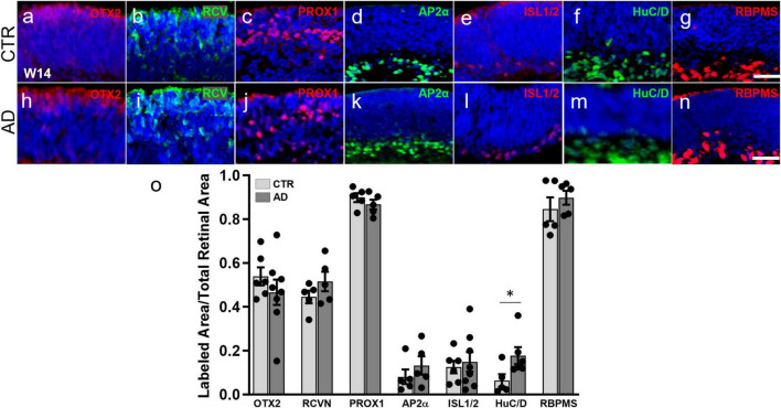
FIGURE 1.
Alzheimer’s disease hiPSC lines produce retinal organoids with major retinal cell types and in a pattern similar to control organoids. (a–n) Immunofluorescence for markers of different retinal cell types at 3 months of differentiation in cryosections of AD and control (CTR) organoids shows that all early born retinal cell types are generated. Markers used were: OTX2 (a,h) and recoverin (RCVN) (b,i), for photoreceptor precursors; PROX1 (c,j) for horizontal and amacrine cells; AP2α (d,k) for amacrine cells; ISL1/2 (e,l) for amacrine and retinal ganglion cells; HuC/D (f,m), for amacrine and ganglion cells; and RBPMS (g,n) for retinal ganglion cells. (o) Immunofluorescence quantification shows no significant differences in OTX2, recoverin, PROX1, AP2α, ISL1/2, and RBPMS. Mann–Whitney test; *p < 0.05; N ≥ 5 organoids per condition including samples from UCSD239iAPP2-1 and HVRDi001-A-1 (AD lines), and A18945 and ILC42.3 (control lines). Scale bars, 50 μm. Bars represent mean ± SEM (error bars).

