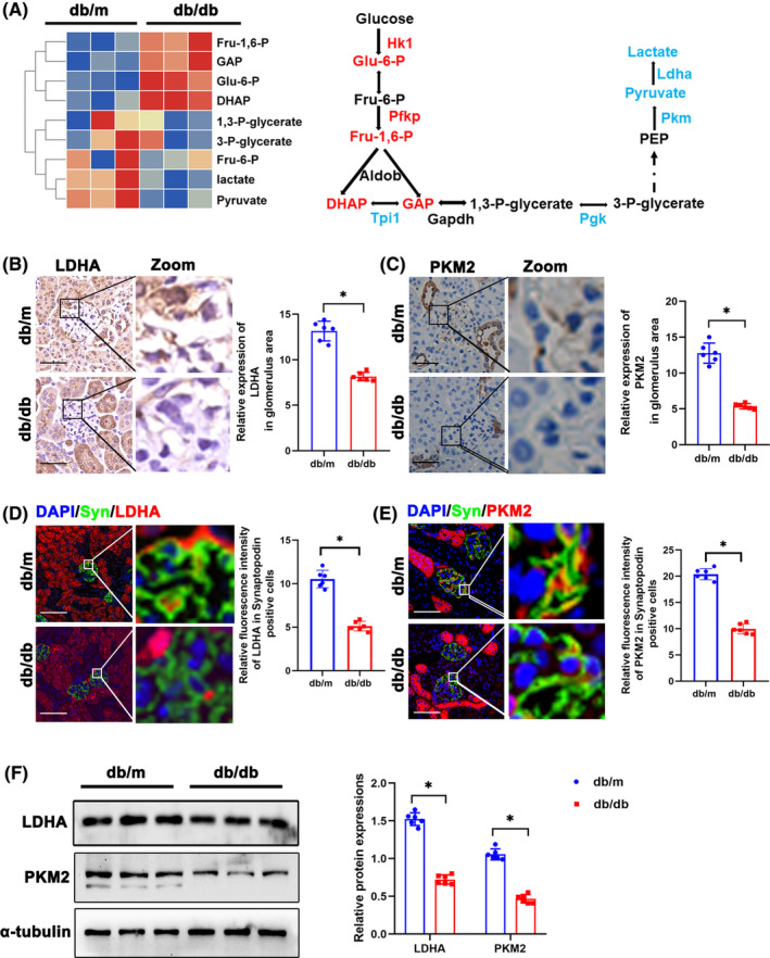
FIGURE 1.

Glycolysis dysfunction and accumulation of DHAP in glomeruli of diabetic mice. (A)Metabolomics of glomeruli of db/m and db/db mice. Altered metabolites in glycolysis were showed in heat map. The schematic flow illustrates the representative genes and metabolites in glycolysis cascade. Letters in red represent upregulated genes and ones in blue represent downregulated metabolites from glomeruli of db/db mice in comparison to db/m mice. (B) Representative immunohistochemistry staining of glomerular LDHA in db/m and db/db mice. (C) Representative immunohistochemistry staining of glomerular PKM2 in db/m and db/db mice. (D) Representative immunofluorescent staining of LDHA in podocyte of glomeruli from db/m and db/db mice. Synaptopodin was used as podocyte markers. (E) Representative immunofluorescent staining of PKM2 in podocyte of glomeruli from db/m and db/db mice. Synaptopodin was used as podocyte markers. (F) Western blotting analysis of the expression of LDHA and PKM2 in glomeruli of db/m and db/db mice. α‐tubulin was used as loading control. Scale bars: 20 μm. n = 6. *p < 0.05. 1,3‐P‐glycerate, 1,3‐bisphosphoglycerate; 3‐P‐glycerate, 3‐phosphoglycerate; Aldob, aldolase B type; DHAP, dihydroxyacetone phosphate; Eno1, enolase 1; Fru 6‐P, D‐fructose 6‐phosphate; Fru‐1,6‐P, fructose 1, 6‐bisphosphate; GAP, Glyceraldehyde 3 phosphate; Gapdh, glyceraldehyde 3‐phosphate dehydrogenase; Glo1, glyoxalase 1; Hagh, hydroxyacyl glutathione hydrolase; Hk, hexokinase; Ldh, lactate dehydrogenase; ns, not significant; Pdh, pyruvate dehydrogenase; PEP, Phosphoenolpyruvic acid; Pfk, phosphofructokinase; Pgk, phosphoglycerate kinase; Pgm1, phosphoglucomutase‐1; Pkm, pyruvate kinase isoenzyme; PKM2, pyruvate kinase isoenzyme 2; Tpi1, triosephosphate isomerase 1.
