FIGURE 6.

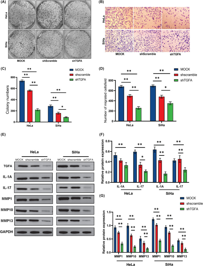
Functional validation of TGFA in CESC cell lines. (A, C) The proliferation of HeLa and SiHa cells was detected by colony formation assay. (B, D) The invasion ability of HeLa and SiHa cells was detected by Transwell assay. (E–G) The expression of IL‐1A, IL‐17, MMP1, MMP10 and MMP13 after TGFA expression reduction was detected by western blot. Significance identifier: ns (no significance), p ≥ 0.05; *, p < 0.05; **p < 0.01; ***p < 0.001.
