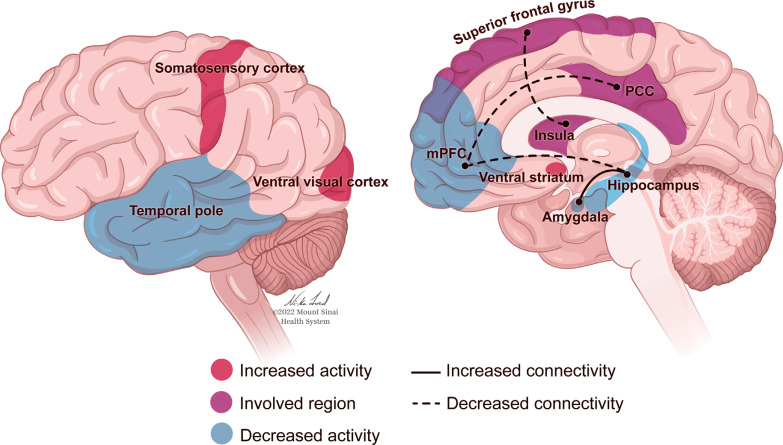
Fig. (4).
Effects of MDMA on the brain. MDMA reduces cerebral blood flow in the right amygdala and hippocampus [376]. fMRI findings show that MDMA attenuates amygdala reactivity in response to participant exposure to angry faces while amplifying ventral striatum response to happy faces [365]. The left anterior temporal cortex, a region proximal to and densely connected with the amygdala, demonstrates reduced activation in healthy participants following MDMA intake. This reduced activity occurred while participants were reflecting upon their worst autobiographical memories and was correlated with a reportedly less negative subjective experience of these memories. In contrast, participants reported their favorite autobiographical memories as more emotionally intense and positive after MDMA, which correlated with increased activations of the ventral visual and somatosensory cortices [377]. Resting-state functional connectivity (RSFC) between the ventromedial prefrontal cortex and posterior cingulate cortex is attenuated following MDMA consumption, an observation that has also been found following psilocybin administration [376, 378, 379]. Patients with PTSD demonstrate increased RSFC between the medial prefrontal cortex and hippocampus [375], whereas MDMA decreases RSFC between these two regions [376]. Additionally, increases in amygdala-hippocampal RSFC were observed after MDMA administration [376]; a notable finding as decreased amygdala-hippocampal RSFC has been seen in patients with PTSD [142]. Lastly, MDMA has been shown to decrease Salience Network FC, specifically between the right insula and superior frontal gyrus [380].

