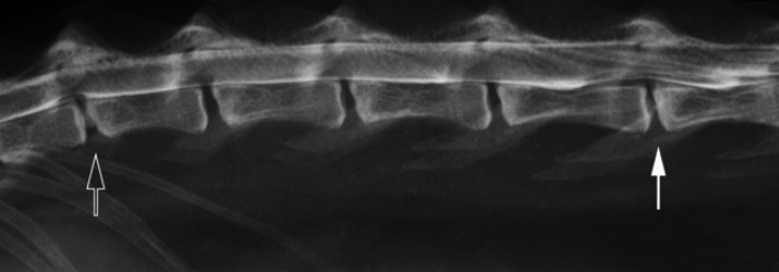
Fig 2.
Right lateral view of the myelogram of Cat 1. Mineralisation of the T13-L1 disc is apparent, combined with a ventral extradural spinal cord compression at that site (open arrow). A broad ventral extradural compression is seen at L4–5, with narrowing of that disc space and sclerosis of the vertebral endplates (closed arrow).

