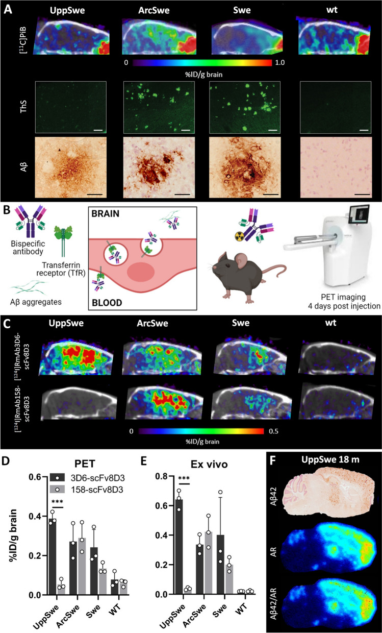
Fig. 3.
PET imaging. A Sagittal [11C]PiB-PET/CT images of 18- months-old tg-UppSwe mice in comparison with tg-ArcSwe, tg-Swe and wt mice, acquired 40–60 min after [11C]PiB injection, with corresponding post mortem brain tissue stained with thioflavin S (ThS, scale bar: 200 µm) and Aβ42 immunostaining (scale bar: 50 µm) below. B Bispecific Aβ antibody undergoing transcytosis across the BBB endothelium. Radiolabeled bispecific Aβ antibody ligands were used for in vivo immunoPET imaging four days after injection. C ImmunoPET/CT images of 18-month-old tg-UppSwe, tg-ArcSwe, tg-Swe and wt mice, injected with [124I]RmAb3D6-scFv8D3 or [124I]RmAb158-scFv8D3. D Quantification of immunoPET data in groups of mice (n = 3 per group). E Ex vivo quantification of brain radioactivity in the same mice as (D). F Aβ42 immuno-staining, ex vivo autoradiography (AR) and an Aβ42/AR merged image of brain sections from tg-UppSwe mouse injected with [124I]RmAb3D6-scFv8D3. ***P < 0.01

