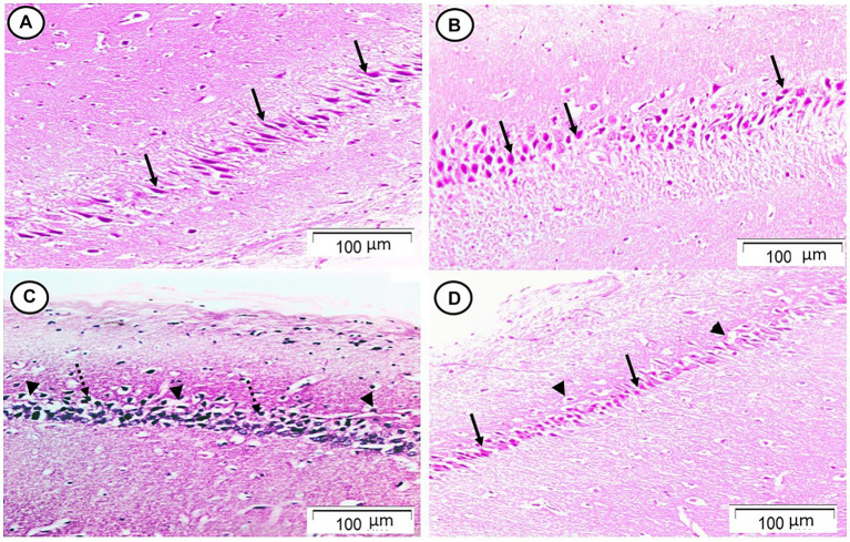
Figure 1.
Photomicrograph of hippocampal cornu ammonis region 1 (CA1). (A,B) The control and Rn groups, respectively, showed the pyramidal cell layer formed of small pyramidal neurons with large vesicular nuclei and notable nucleoli (arrows). (C) The Sco group showed that most of the pyramidal neurons appeared to be deeply stained with pyknotic nuclei (dotted arrows). There were some halos that indicated neuronal loss (arrow heads). (D) The Rn + Sco group showed that most of the pyramidal neurons appeared normal (arrows). However, there were halos indicating neuronal loss still seen (arrow heads) (H & E, x 200).

