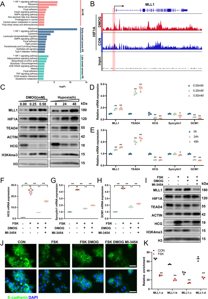
Fig. 6.
HIF1A regulates trophoblast syncytialization through the MLL1/H3K4me3/TEAD4 axis. A KEGG analyses performed on women at different stages of PE and healthy controls. B HIF1A chromatin immunoprecipitation sequencing (ChIP-seq) signals for MLL1 in cells treated with DMOG or DMSO. C Western blots of MLL1, HIF1A, TEAD4, HCG, and H3K4me3 in BeWo cells treated with a gradient of DMOG concentrations (0, 0.25, and 0.50 mM) and hypoxia exposure time (0, 24, and 48 h). D and E mRNA levels of MLL1, TEAD4, and STB markers in BeWo cells treated with a gradient of DMOG concentrations and hypoxia exposure time. F-J mRNA levels of STB markers (F–H), western blots of MLL1, HIF1A, TEAD4, HCG, and H3K4me3 (I) and immunostainings of E-cadherin (green) and DAPI (blue) (J) in BeWo cells treated with FSK (25 μM) in the absence or presence of DMOG (0.25 mM) and MI-3454 (5 μM). Scale bar, 40 μm. K Quantitative ChIP analysis of HIF1A at the MLL1 promoter in FSK-treated BeWo cells. The values are normalized to IgG. Data are presented as the means ± SD. **P < 0.01, *P < 0.05. DMOG, dimethyloxallyl glycine; HIF1A, hypoxia-inducible factor 1A

