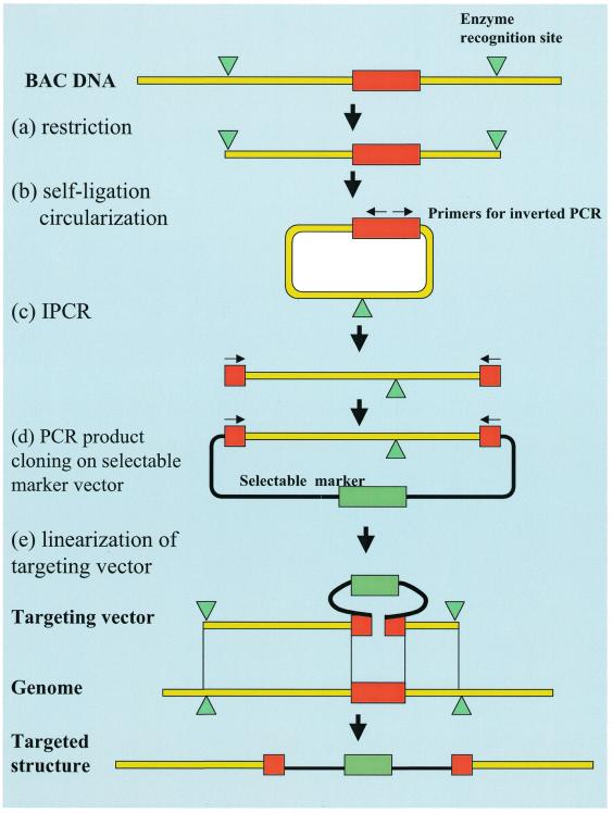
Figure 1.
Procedure for constructing a targeting vector by the IPCR method. (a) An isolated BAC clone which contains the target gene is digested with the proper six-base cutter. The restriction sites are indicated with green triangles and the target sites are indicated with red rectangles. (b) The restricted fragments are circularized by self-ligation. (c) Using the primers designed for IPCR (indicated by arrows) and the circularized DNA as a template, only the target genomic region is amplified. (d) The amplified target region is inserted into the cloning vector with selectable markers (indicated with green rectangles). (e) The linearization of the targeting vector is ready for transfection. The homologous recombination between the genomic target and the replacement targeting vector is also shown schematically.

