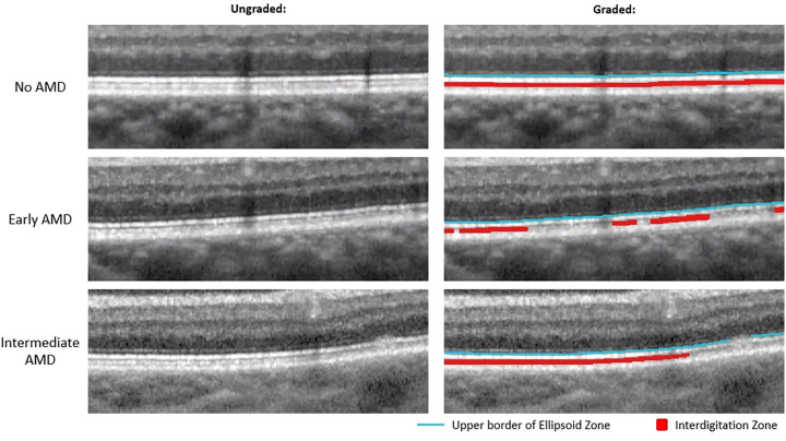
Figure 1.
Magnified view of outer retinal bands in no AMD, early AMD, and intermediate AMD eyes. Left: Unaltered magnified OCT B-scans. Right: Same magnified OCT B-scan as the left side but graded for EZ and IZ. The blue line is the upper border of the EZ which was used as a proxy for discernable EZ, and the red area represents discernable IZ.

