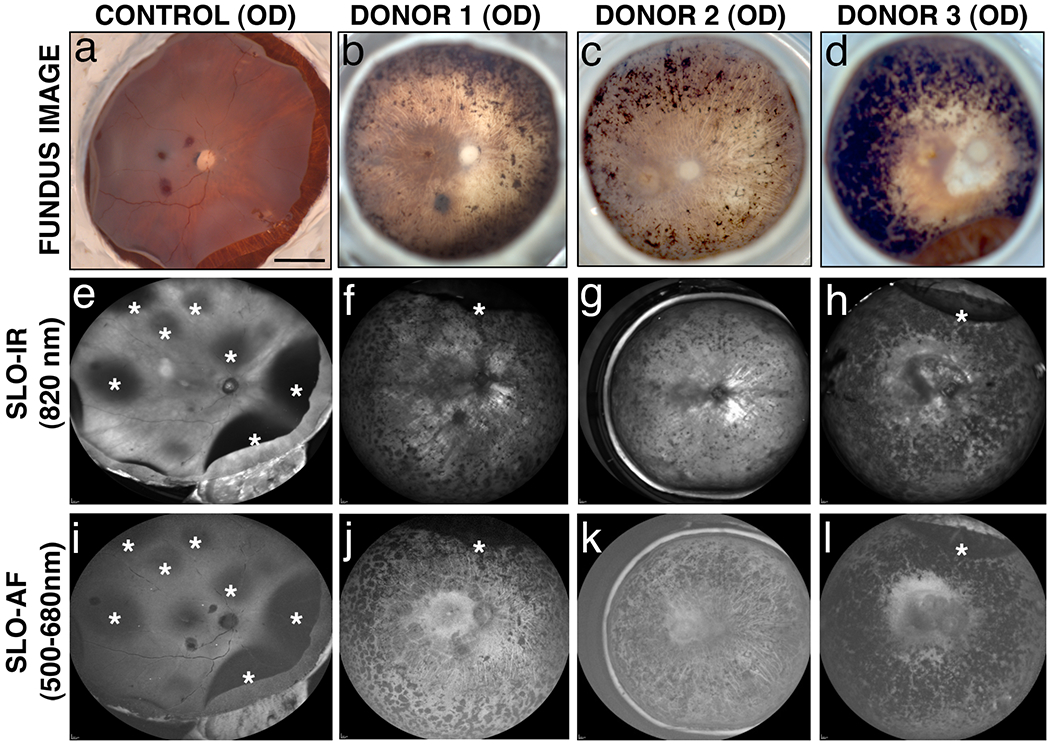
Fig. 2. Ex-vivo imaging of arRP donor eyes with EYS mutations.

Fundus images (a-d) and SLO images (e-l) were collected from donor 1, 2 and 3 and an age-similar control. In the control eye, detached retinas are apparent with all imaging modalities (a, e, i, *). In all three arRP eyes, fundus images reveal bone spicule pigment in mid-peripheral and peripheral areas to varying degrees (b-d). SLO-IR imaging identified degeneration in the entire posterior pole region including the macula, perimacula and areas surrounding the optic nerve due to focal loss of RPE in donors 2 (g) and 3 (h) as compared to an age-matched control eye (e). SLO-AF imaging identified hypofluorescence in one contiguous region involving the macula and area surrounding the optic disk of donor 3 (l) as opposed to the individually demarcated and isolated regions seen with both donors 1 (j) and 2 (k) and the control eye (i). Scale bars in fundus image = 0.5 cm.
