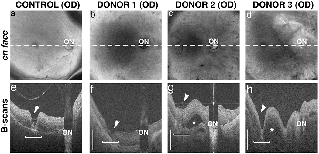
Fig. 3. Ex-vivo OCT imaging of arRP donor eyes with EYS mutations.

OCT images were collected from donors 1, 2 and 3 and an age-matched control. En face images reveal the location (dashed lines) of the in-depth, B-scan images of control (a) and arRP donors 1 (b), 2 (c) and 3 (d). The fovea (arrowhead) and optic nerve (ON) were identified in all donor eyes. In-depth B-scans from the control eye (e) revealed a normal appearing retina with clearly defined fovea, some evidence of laminar architecture, and no appreciable evidence of retinal thinning or degeneration. Images from the arRP donors (f-h) revealed retinas of appreciable thickness but with less organized architecture and integrity in the macular region (horizontal bracket). In contrast, the perimacular regions showed some evidence of thinned retina relative to the control retina suggesting degeneration. Donor’s 2 (g) and 3 (h) had macular (*) and choroidal (arrow) detachments as indicated in the B-scans. B-scan scale is 0.5mm.
