
Fig. 6. Immunocytochemistry of arRP retinal sections with EYS mutations stained with cone-specific antibodies.
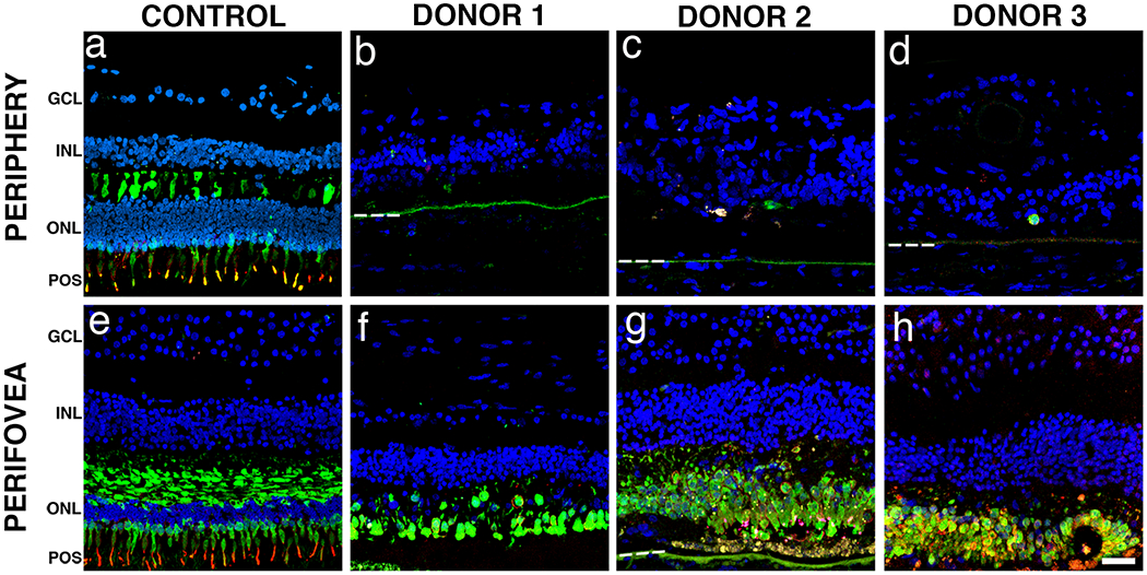
Immunofluorescence of arRP retinal sections labeled with antibodies to cone arrestin (green) and red/green cone opsin (red) showed significantly decreased staining in the periphery when compared to control. In the peripheral (a) and perifoveal (e) regions of the control retina, cone arrestin was distributed along the entire plasma membrane, from the tip of the outer segment to the synaptic base, while the red/green cone opsin was restricted to the outer segments. Cone-specific labeled cells were mostly absent from the periphery of all three arRP donor retinas (b-d). In contrast, cone-specific labeled cells were present but highly disorganized in the perifovea of donor 1 (f). Interestingly, the perifovea from both donor 2 (g) and 3 (h) displayed disorganized cone-specific labeled cells concentrated in the areas that still maintained RPE. Bruch’s membrane is indicated by hashed white line. Nuclei were labeled with TO-PRO-3. GCL= ganglion cell layer; INL= inner nuclear layer; ONL= outer nuclear layer; POS = photoreceptor outer segments. Scale bar = 40μm.