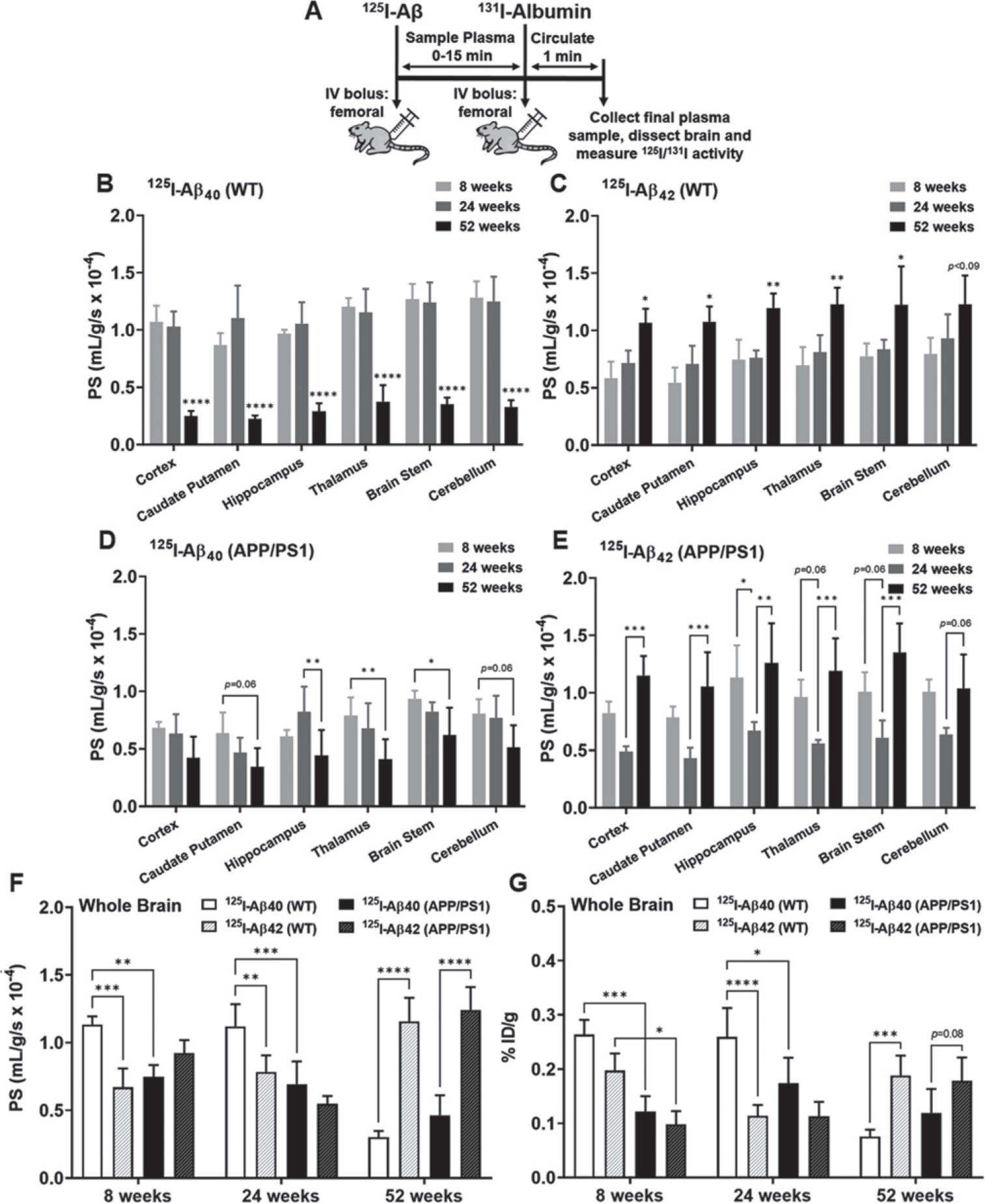
Fig. 2.

Brain uptake of 125I-Aβ40 and 125I-Aβ42 in WT and APP/PS1 mice at 8, 24, and 52 weeks. A) Experiment scheme. WT or APP/PS1 transgenic mice were bolus injected with 100Ci of 125I-Aβ40 or 125I-Aβ42 into the femoral vein. At the end of the experiment, 100μCi of 131I-albumin was injected to serve as a marker of Vp. The brain regions were dissected and assayed for radioactivity. Shown are the PS value estimates for 125I-Aβ40 (B, D) and 125I-Aβ42 (C, E) uptake at various brain regions. F) The PS values are shown for the whole brain. G) The overall brain accumulation was assessed as % ID/g. Data represent mean±SD, n = 3–5. *p < 0.05, **p < 0.01, ***p < 0.001, and ****p < 0.0001; two-way ANOVA with Bonferroni post-tests).
