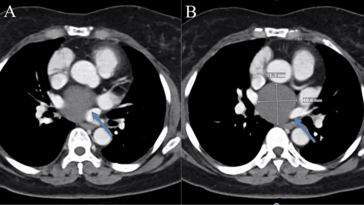
Figure 1. CT scans of the mediastinum and pulmonary hila, showing no lymph node enlargement in the axilla, supraclavicular area, mediastinum, or hilar region. A subcarinal low-density lesion measuring 5.1 × 4.9 cm (A) is observed, which was previously measured as 4.9 × 4.9 cm. No suspicious internal enhancing nodules are detected (B).
CT, computed tomography

