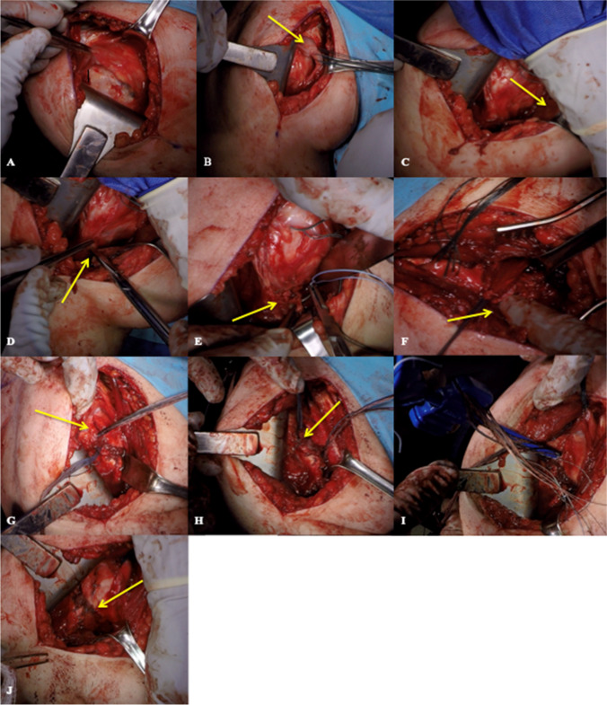
Fig. 2.
Open, anterior LD tendon transfer. A Exposure to the bare lesser tuberosity. B Torn subscapularis retrieved and elevated to accommodate tendon transfer. C Pectoralis retracted away from LD tendon. D LD tendon carefully dissected away from teres major. E Traction sutures placed LD tendon. F Gentle digital mobilization achieves adequate excursion to transfer LD tendon to lesser tuberosity. G Triple-loaded all tape suture placed at inferior aspect of lesser tuberosity. H Traction suture through LD tendon tied into native supraspinatus anterior cable. I Triple-loaded all tape sutures passed through 5.5-mm SwiveLock anchor, placed superior to lesser tuberosity, providing broad footprint reduction. J Remnant subscapularis sutured over the LD tendon, providing additional biologic support

