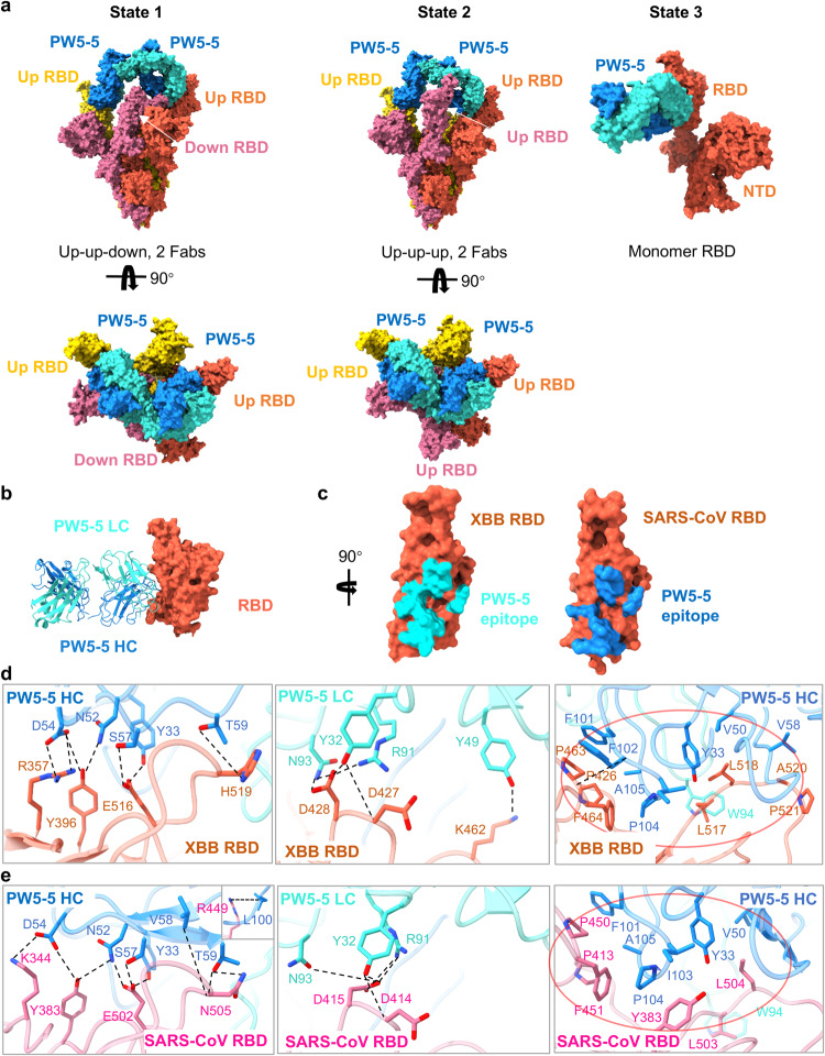
Fig. 5. Cryo-EM structure of PW5-5 IgG with Omicron XBB and SARS-CoV S trimer.
a Cryo-EM structures of the XBB S in complex with the antibody PW5-5 IgG. b Structure of XBB RBD–PW5-5. The RBD is displayed in tomato. The heavy chain and light chain of PW5-5 are shown as ribbons colored in dark blue and light blue, respectively. c Close-up view of the PW5-535 epitope on XBB and SARS-CoV RBD. d Detailed interactions between the PW5-5 and XBB S RBD. e Detailed interactions between the PW5-5 and SARS-CoV S RBD. Hydrogen bonds are represented by dashed lines. Hydrophobic interactions are marked with red circles.

