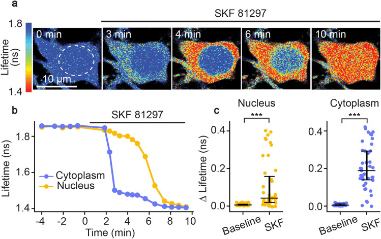
Figure 3.
Functional response of D1R-SPNs to D1R activation in acute slices of Tg(Drd1a-Cre); FLIM-AKARflox/flox mice. (a) Time-lapse heatmaps of 2pFLIM images of an example SPN in acute slices of the dorsal striatum in response to the D1R agonist SKF 81297 (1 µM). Dotted line indicates the location of nucleus. (b) Example traces of fluorescence lifetime response of the D1R-SPN shown in (a). (c) Quantification of change of fluorescence lifetime in response to SKF 81297 in D1R-SPNs (n = 43 cells from 6 mice). Data are represented as median with 25th and 75th percentiles (***p < 0.001, Wilcoxon signed rank test).

