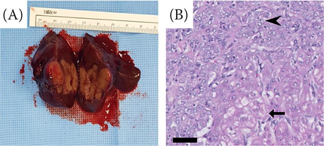
Figure 2. Gross morphological and histopathological features of hepatic mass in a dog with mixed HCC-CC.

(A) Note the multiple nodules within the massive hepatic tumour. (B) Histopathology of hepatic mass revealed that it was composed of both neoplastic hepatocytes and the biliary epithelium. Neoplastic hepatocytes contain abundant vacuolated cytoplasm and irregular oval nuclei with small nucleoli (arrow). The neoplastic biliary epithelium is cuboidal or polygonal with eosinophilic cytoplasm and irregular oval nuclei with variably prominent nucleoli (arrowhead). Haematoxylin & eosin (H&E) stain; magnification 400 ×; scale bar = 50 μm
HCC-CC = hepatocellular carcinoma-cholangiocarcinoma
