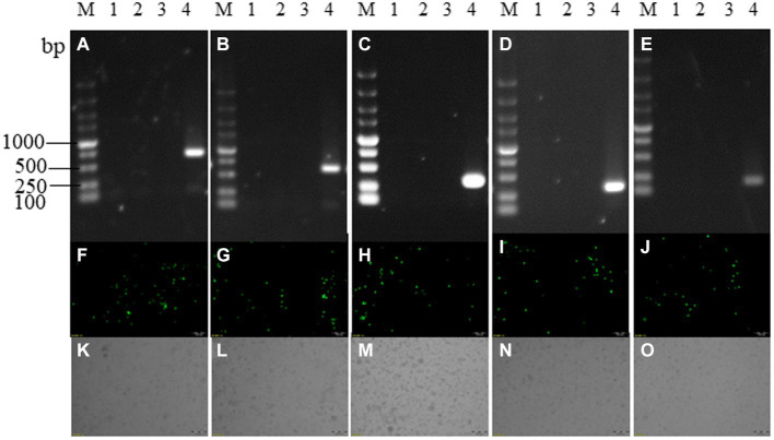
Figure 3.
Recombinant virus identification. Panels (A–E) are PCR identification and purification results of SY18ΔL7L, SY18ΔL8L, SY18ΔL9R, SY18ΔL10L, SY18ΔL11L, panels (F–J) are the fluorescence maps of each single-gene deletion viruses, and panels (K–O) are the bright field of each single-gene deletion viruses. M: DL5000 marker; a1~a3, b1~b3, c1~c3, d1~d3, e1~e3: each single-gene deletion virus detection samples; 4: ASFV SY18 control.SY18ΔL7L and SY18ΔL8L amplified a 643 bp fragment in the presence of ASFV SY18; SY18ΔL9R amplified a 280 bp fragment in the presence of ASFV SY18; SY18ΔL10L, and SY18ΔL11L amplified a 200 bp fragment in the presence of ASFV SY18.

