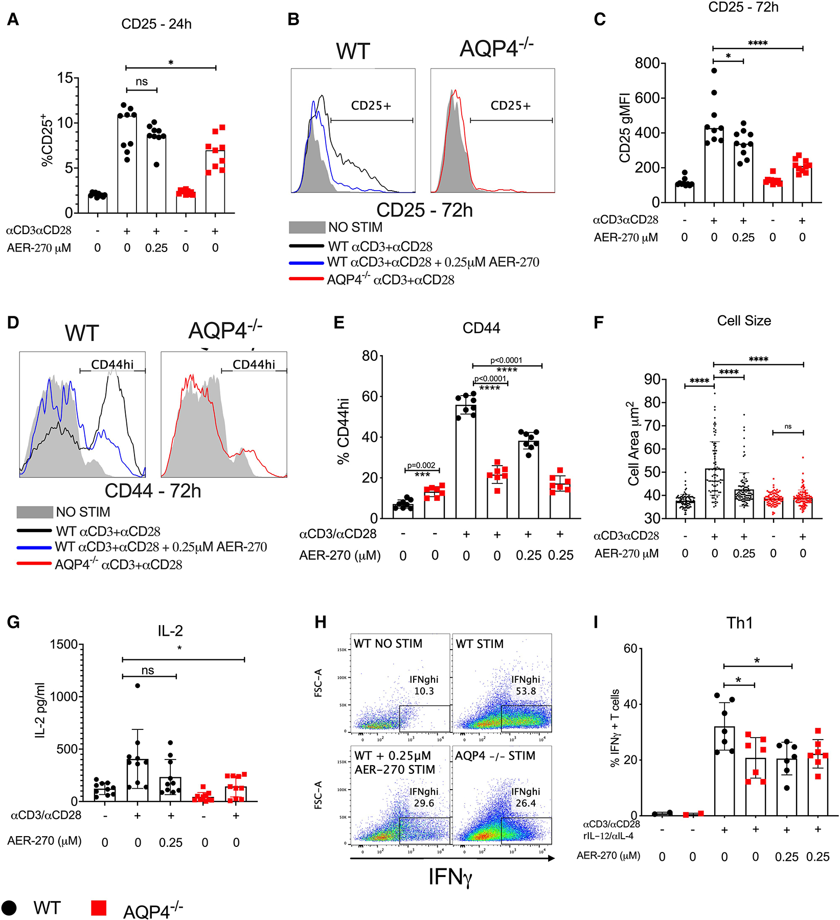
Fig. 1.

Intact AQP4 is required for optimal mouse T cell activation and cytokine production. Mouse WT (●) or AQP4−/− ( ) splenic T cells were stimulated with αCD3/αCD28 antibodies in the presence or absence of 0.25 μM AER-270. (A–C) Expression of early activation marker CD25 was evaluated after 24 h (A) and 72 h (B and C) of stimulation by flow cytometry. (D–G) After 72 h of stimulation, surface activation marker CD44 (D and E) as well as cell size (F) were measured by flow cytometry, and IL-2 was detected from culture supernatants by ELISA from the same stimulation conditions (G). (H–I) WT or AQP4−/− splenic T cells were stimulated for 72 h with αCD3/αCD28 antibodies with the addition of rIL-12/αIL-4, in the presence or absence of 0.25 μM AER-270 and Th1 polarization was evaluated by intracellular IFNγ detection (H&I) via flowcytometry. Data shown are representative of 3 experiments where n = 2–7. *P < 0.05, **P < 0.01, ***P < 0.001, ****P < 0.0001 and ns—P > 0.05 via a 1-tailed Student’s t-test.
